Performance Evaluation of the uEXPLORER Total-Body PET/CT Scanner Based on NEMA NU 2-2018 with Additional Tests to Characterize PET Scanners with a Long Axial Field of View
- PMID: 33008932
- PMCID: PMC8729871
- DOI: 10.2967/jnumed.120.250597
Performance Evaluation of the uEXPLORER Total-Body PET/CT Scanner Based on NEMA NU 2-2018 with Additional Tests to Characterize PET Scanners with a Long Axial Field of View
Abstract
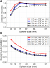
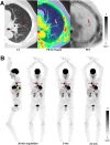
The world's first total-body PET scanner with an axial field of view (AFOV) of 194 cm is now in clinical and research use at our institution. The uEXPLORER PET/CT system is the first commercially available total-body PET scanner. Here we present a detailed physical characterization of this scanner based on National Electrical Manufacturers Association (NEMA) NU 2-2018 along with a new set of measurements devised to appropriately characterize the total-body AFOV. Methods: Sensitivity, count-rate performance, time-of-flight resolution, spatial resolution, and image quality were evaluated following the NEMA NU 2-2018 protocol. Additional measurements of sensitivity and count-rate capabilities more representative of total-body imaging were performed using extended-geometry phantoms based on the world-average human height (∼165 cm). Lastly, image quality throughout the long AFOV was assessed with the NEMA image quality (IQ) phantom imaged at 5 axial positions and over a range of expected total-body PET imaging conditions (low dose, delayed imaging, short scan duration). Results: Our performance evaluation demonstrated that the scanner provides a very high sensitivity of 174 kcps/MBq, a count-rate performance with a peak noise-equivalent count rate of approximately 2 Mcps for total-body imaging, and good spatial resolution capabilities for human imaging (≤3.0 mm in full width at half maximum near the center of the AFOV). Excellent IQ, excellent contrast recovery, and low noise properties were illustrated across the AFOV in both NEMA IQ phantom evaluations and human imaging examples. Conclusion: In addition to standard NEMA NU 2-2018 characterization, a new set of measurements based on extending NEMA NU 2-2018 phantoms and experiments was devised to characterize the physical performance of the first total-body PET system. The rationale for these extended measurements was evident from differences in sensitivity, count-rate-activity relationships, and noise-equivalent count-rate limits imposed by differences in dead time and randoms fraction between the NEMA NU 2 70-cm phantoms and the more representative total-body imaging phantoms. Overall, the uEXPLORER PET system provides ultra-high sensitivity that supports excellent spatial resolution and IQ throughout the field of view in both phantom and human imaging.
Keywords: EXPLORER; PET; performance evaluation; total-body imaging.
© 2021 by the Society of Nuclear Medicine and Molecular Imaging.
Figures


References
-
- Poon JK. The Performance Limits of Long Axial Field of View PET Scanners. Thesis. University of California, Davis; 2013.
-
- Badawi RD, Lewellen TK, Harrison RL, Kohlmyer SG, Vannoy SD. The effect of camera geometry on singles flux, scatter fraction and trues and randoms sensitivity for cylindrical 3D PET: a simulation study. IEEE Trans Nucl Sci. 2000;47:1228–1232.
Publication types
MeSH terms
Grants and funding
LinkOut - more resources
Full Text Sources
Other Literature Sources
Research Materials
