Review
. 2020 Nov;119(5):559-587.
doi: 10.1097/HP.0000000000001346.

Acute Radiation-induced Lung Injury in the Non-human Primate: A Review and Comparison of Mortality and Co-morbidities Using Models of Partial-body Irradiation with Marginal Bone Marrow Sparing and Whole Thorax Lung Irradiation

Affiliations
Review

Acute Radiation-induced Lung Injury in the Non-human Primate: A Review and Comparison of Mortality and Co-morbidities Using Models of Partial-body Irradiation with Marginal Bone Marrow Sparing and Whole Thorax Lung Irradiation

Thomas J MacVittie et al. Health Phys. 2020 Nov.

Abstract

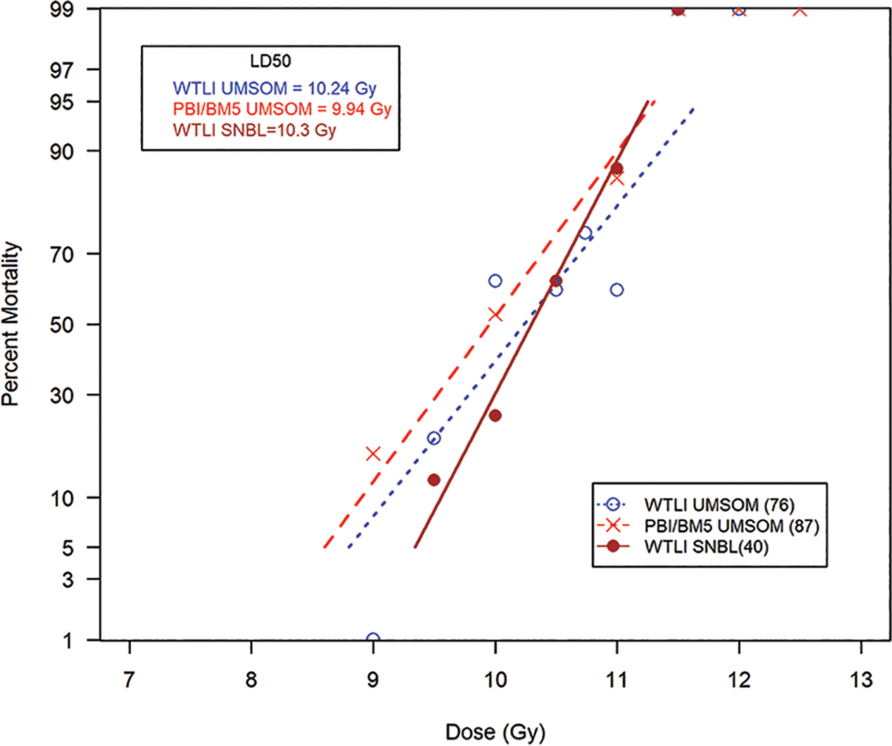
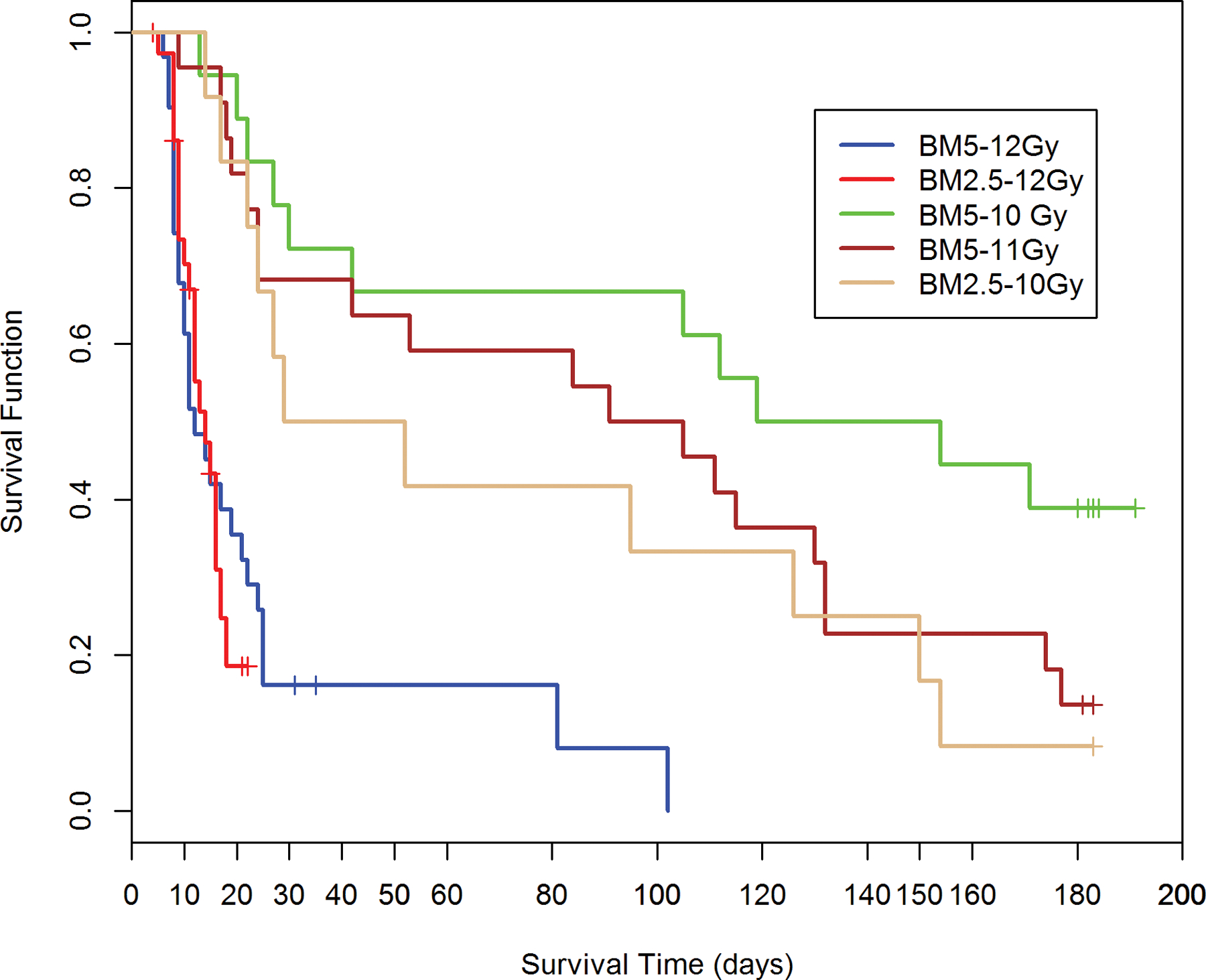
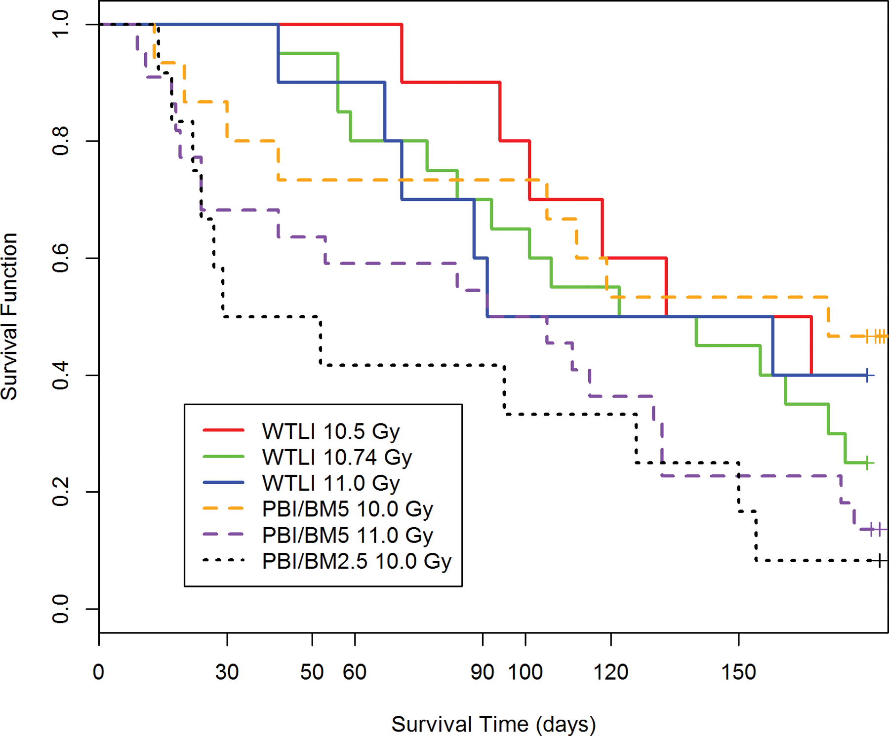
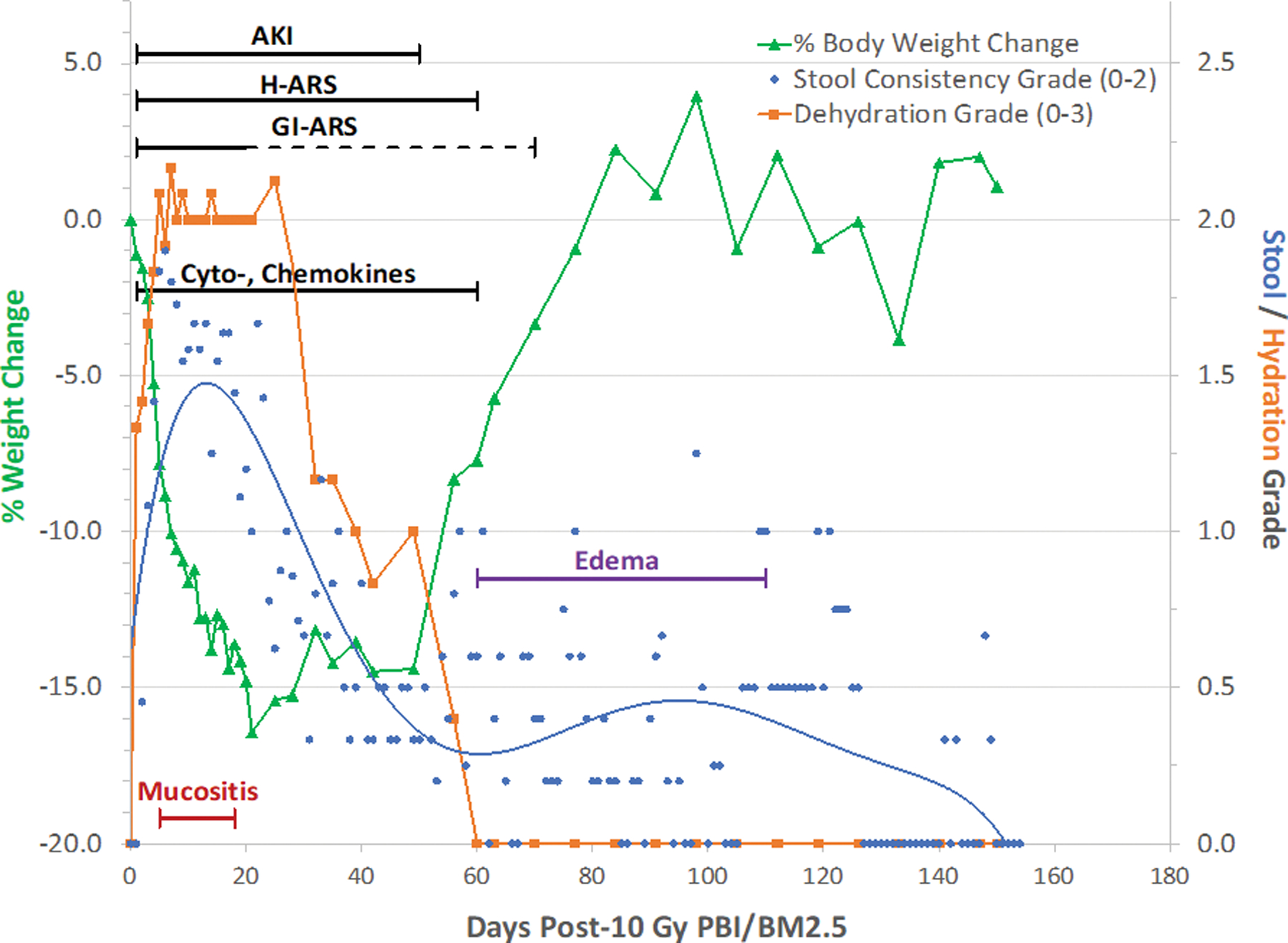
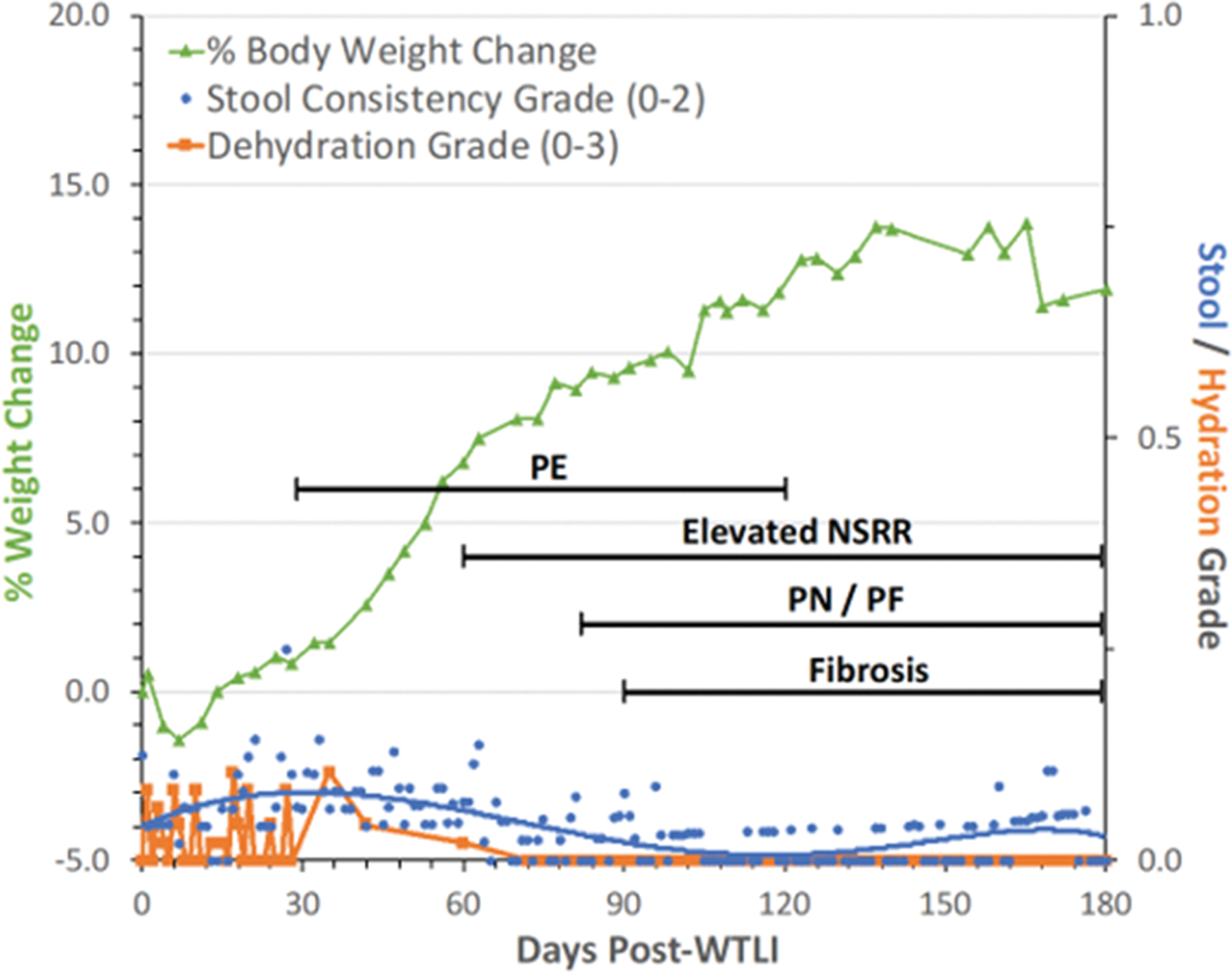
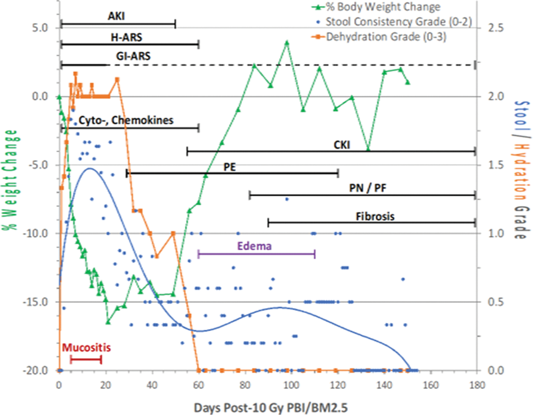
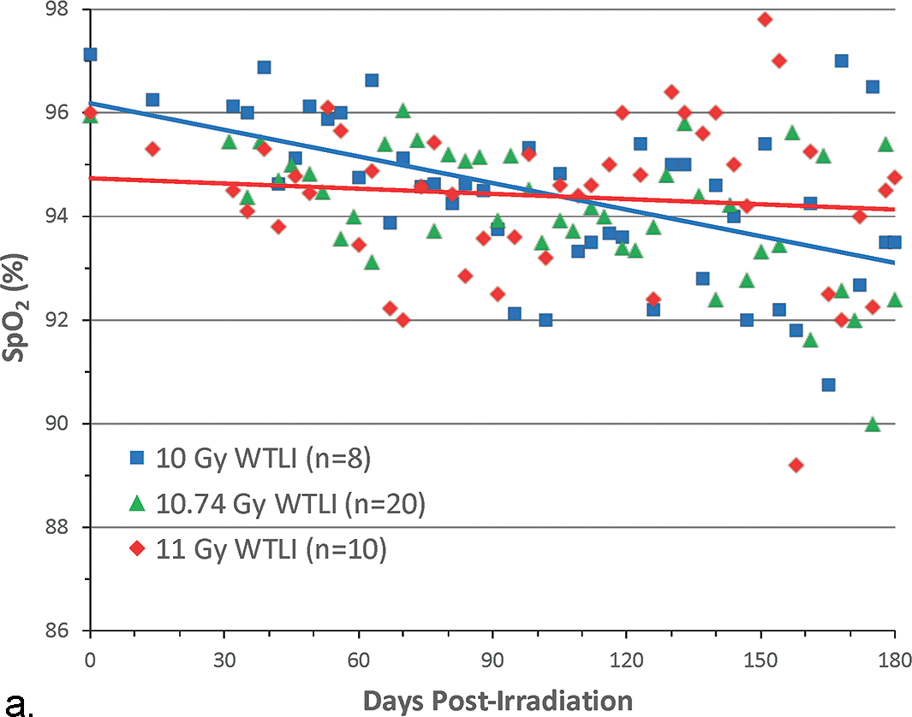
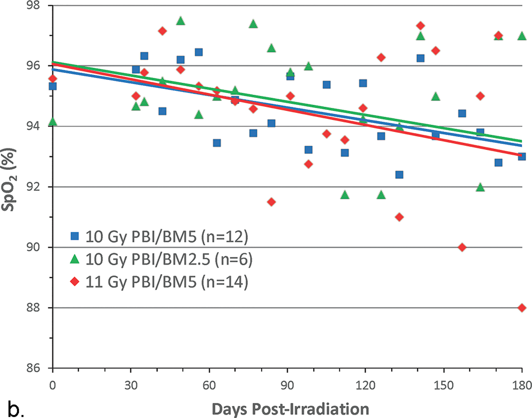
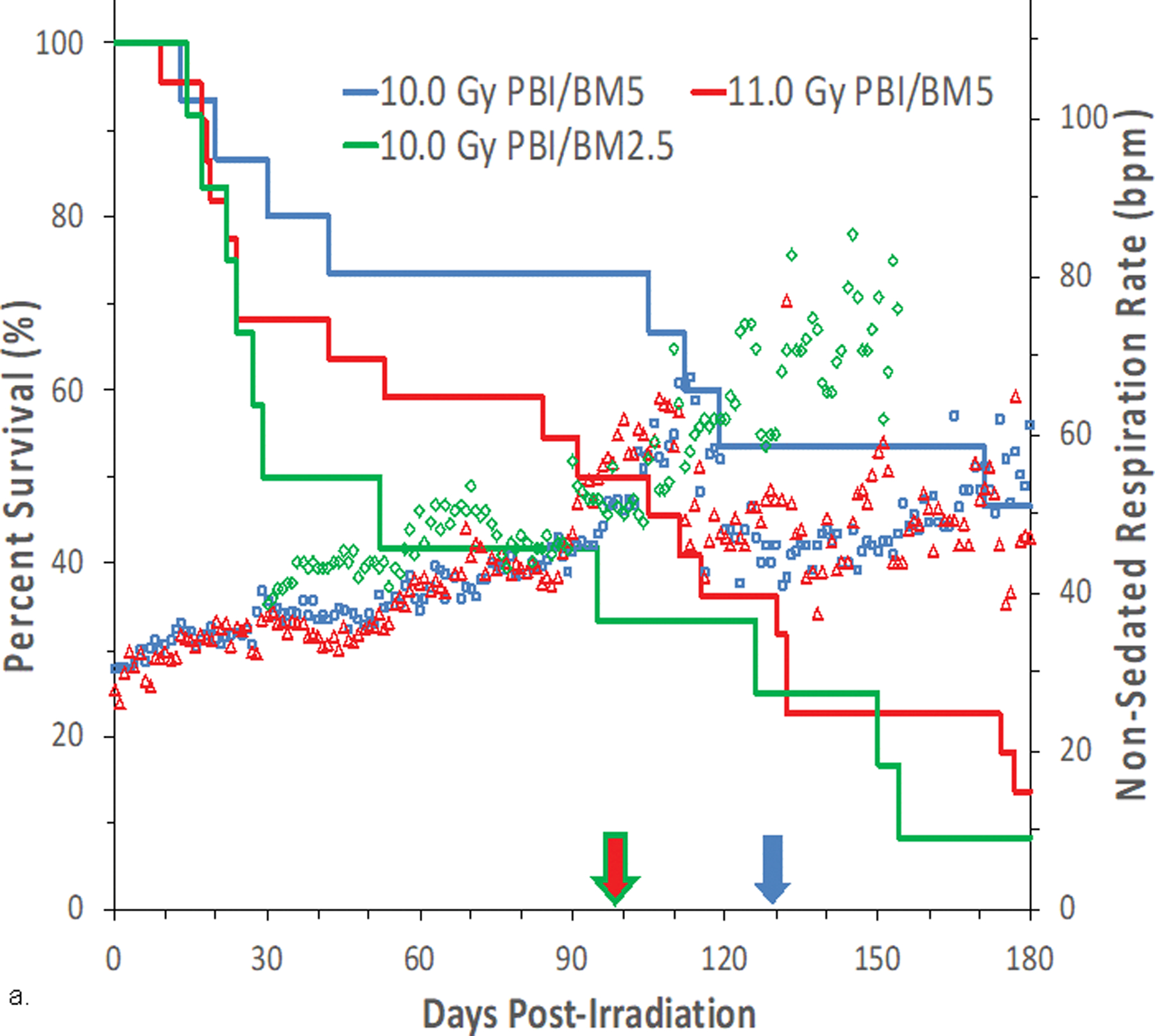
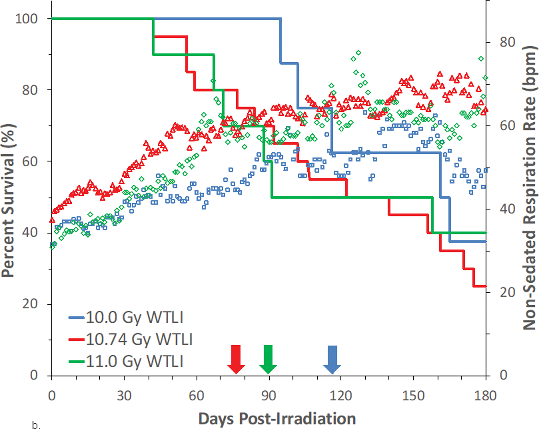
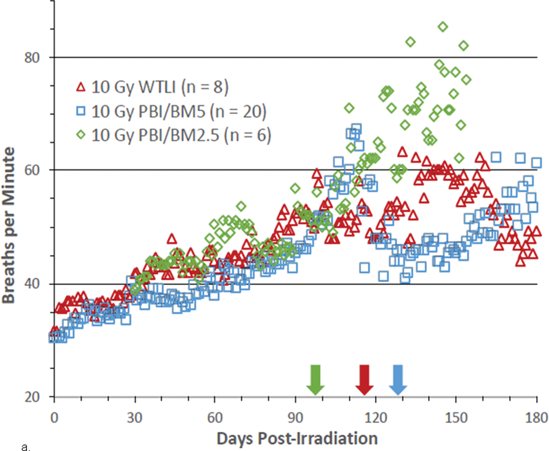
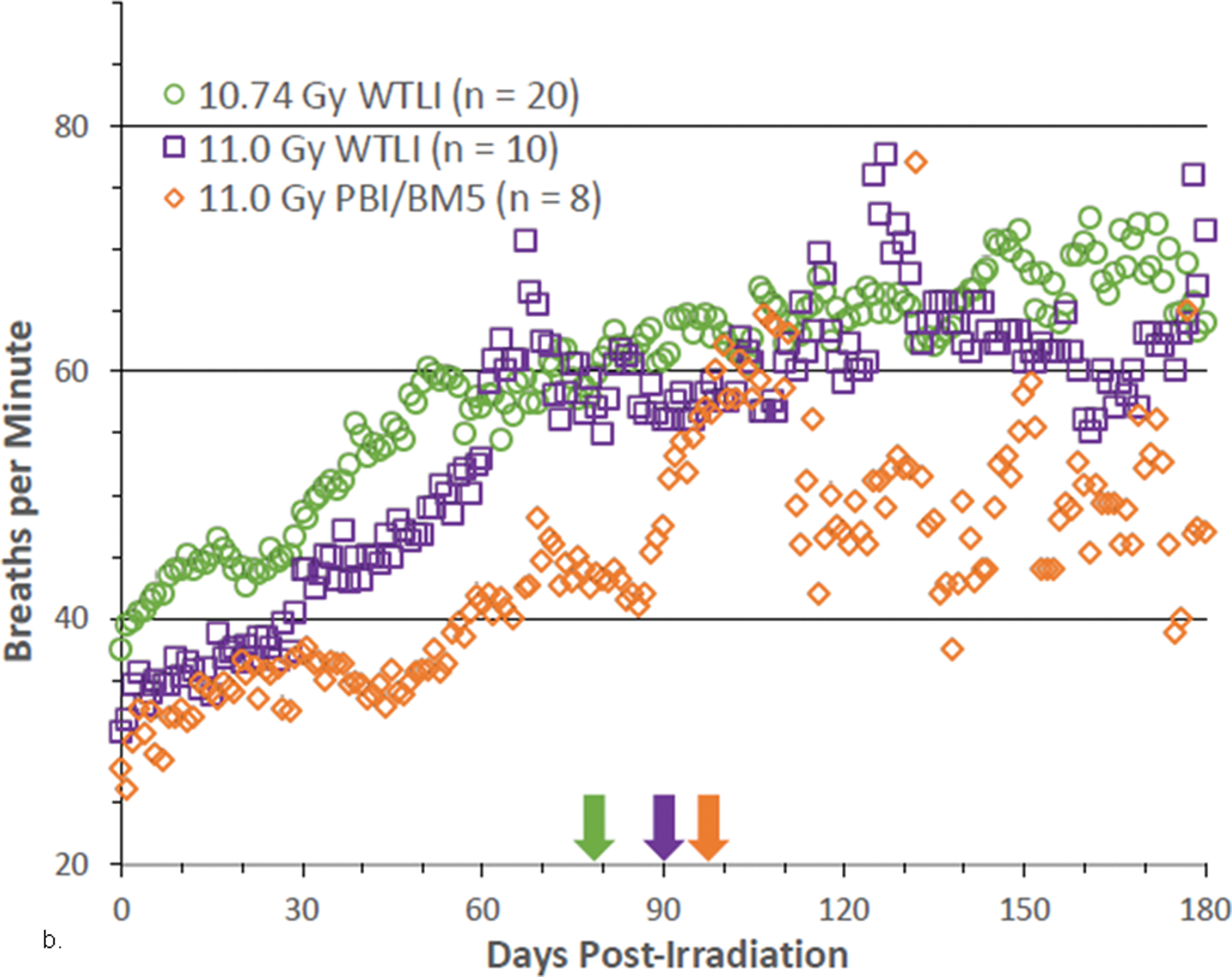
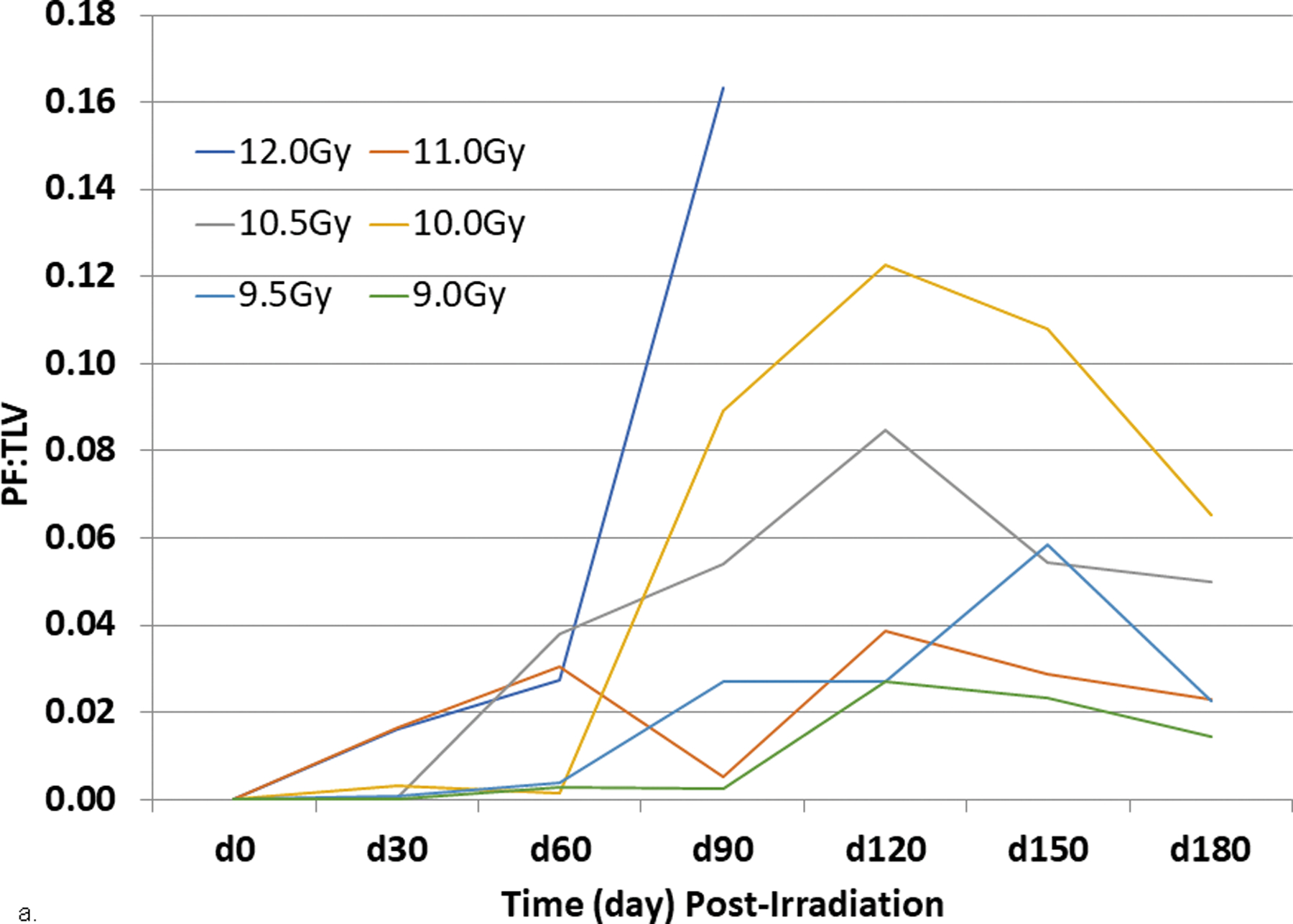
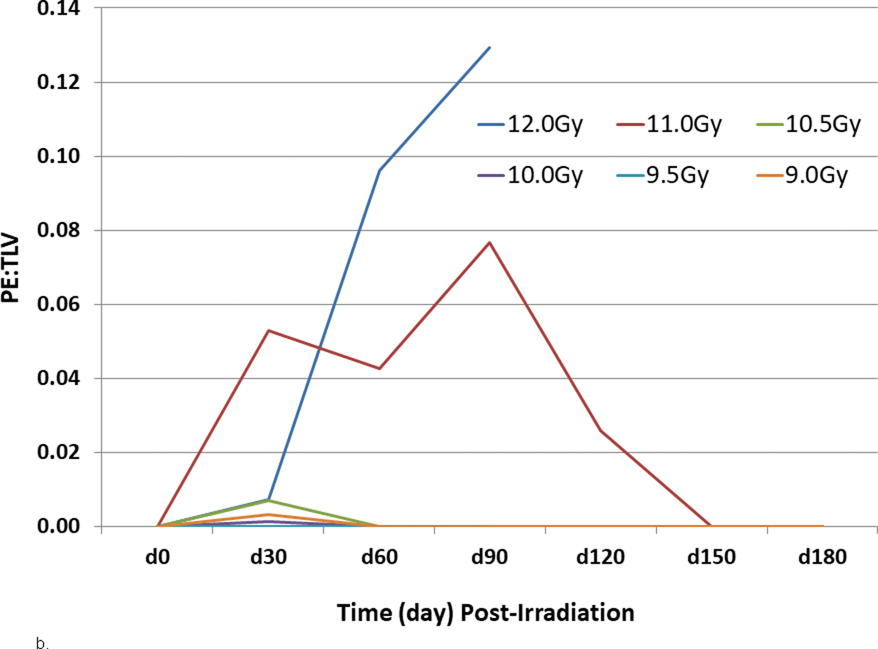
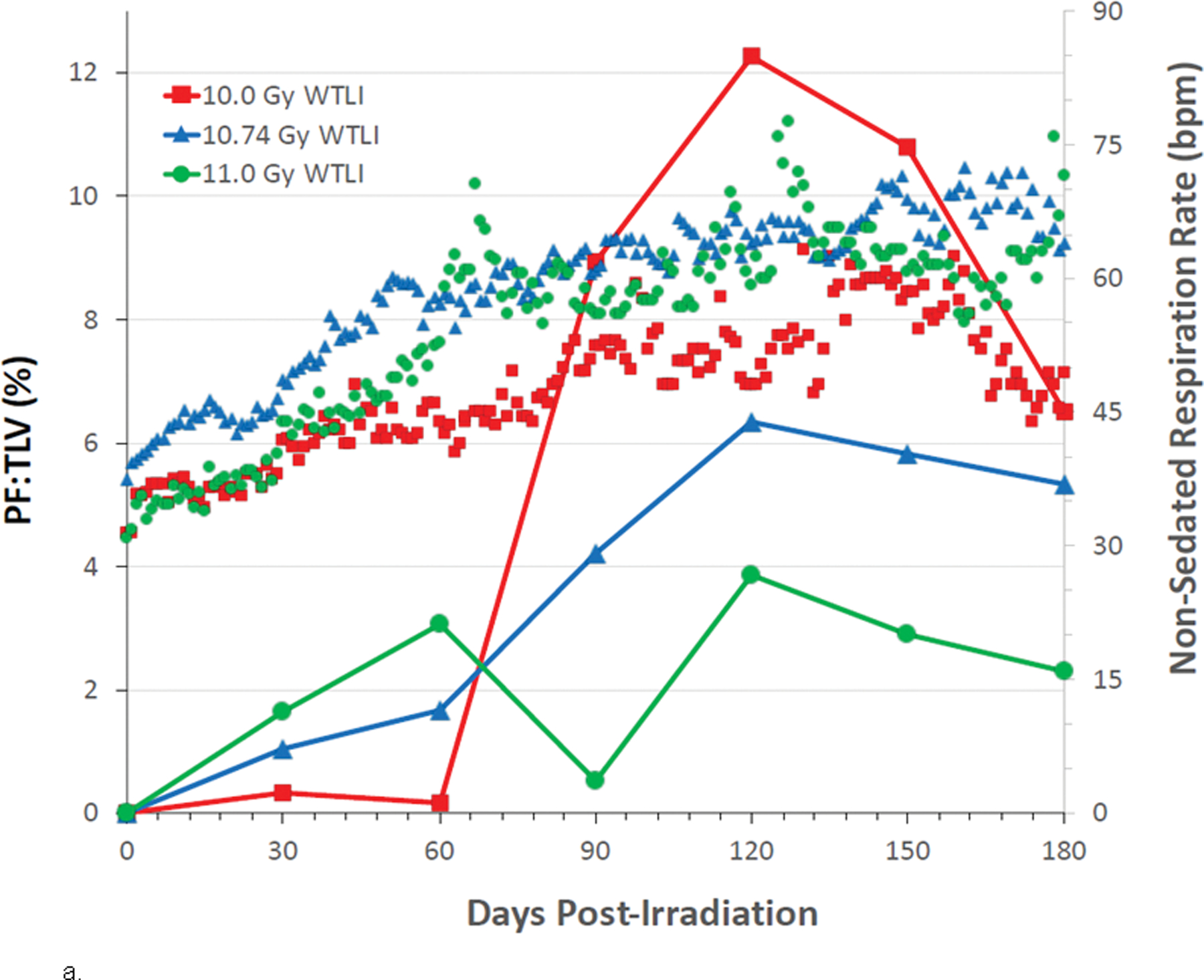
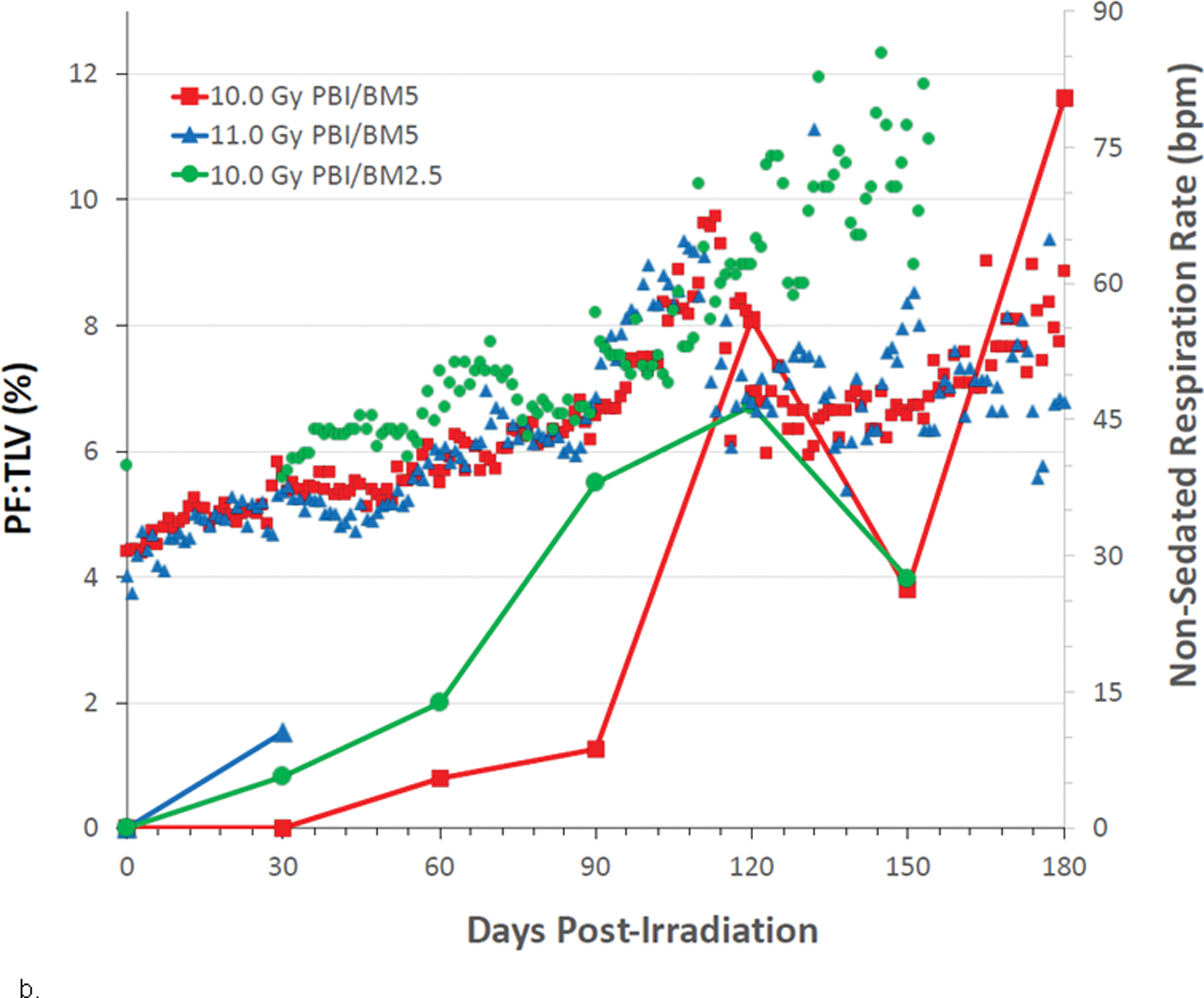
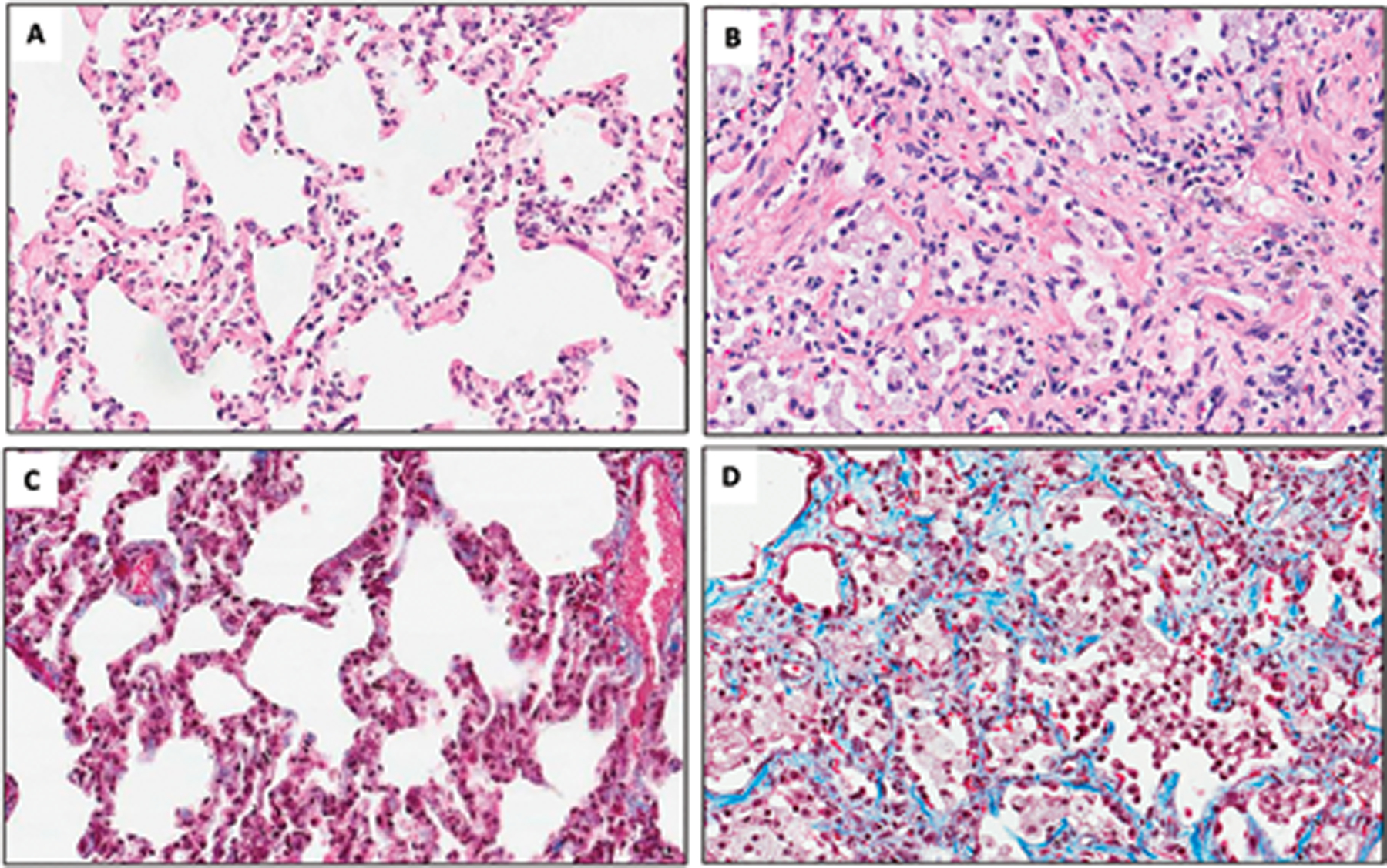
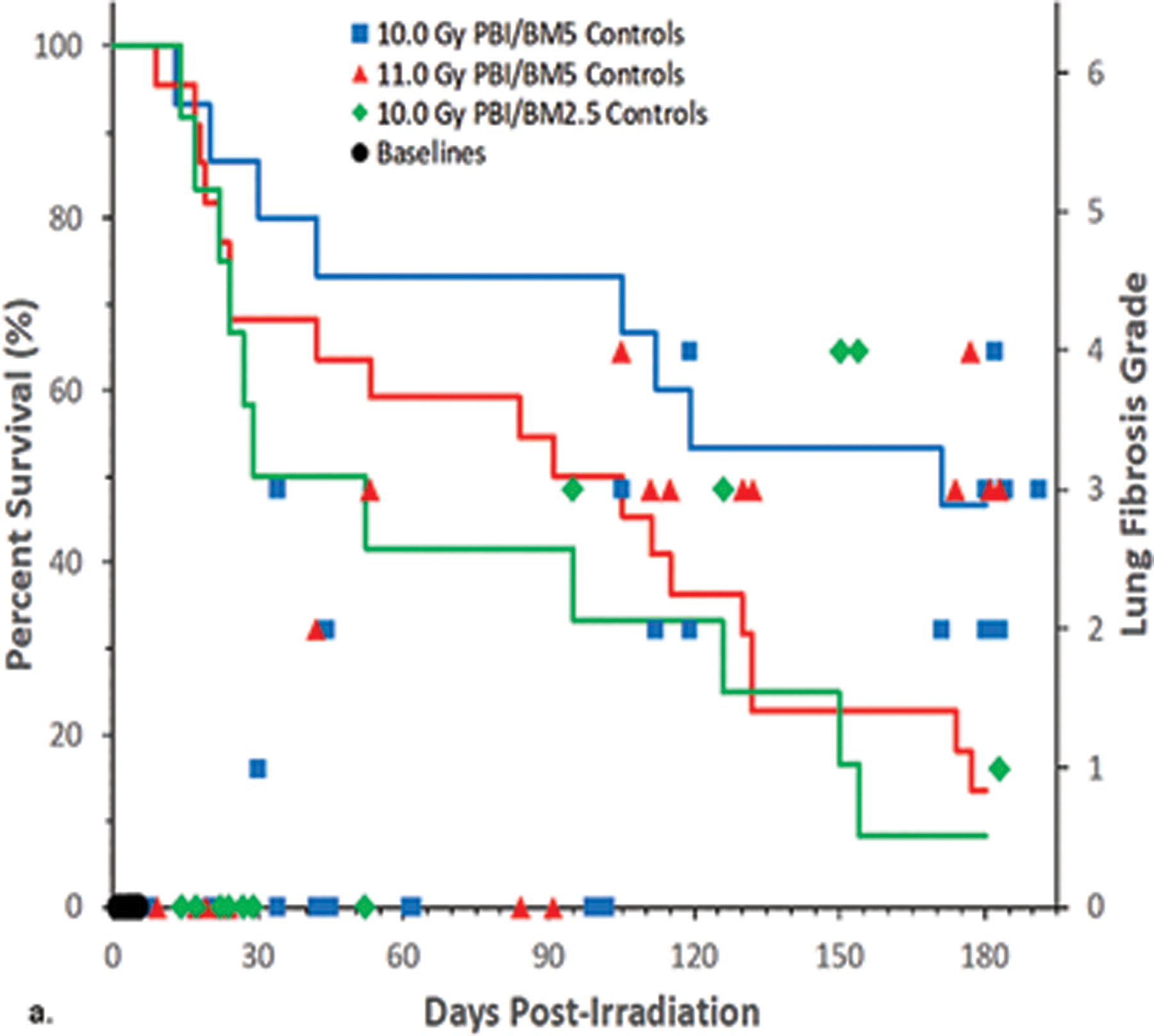
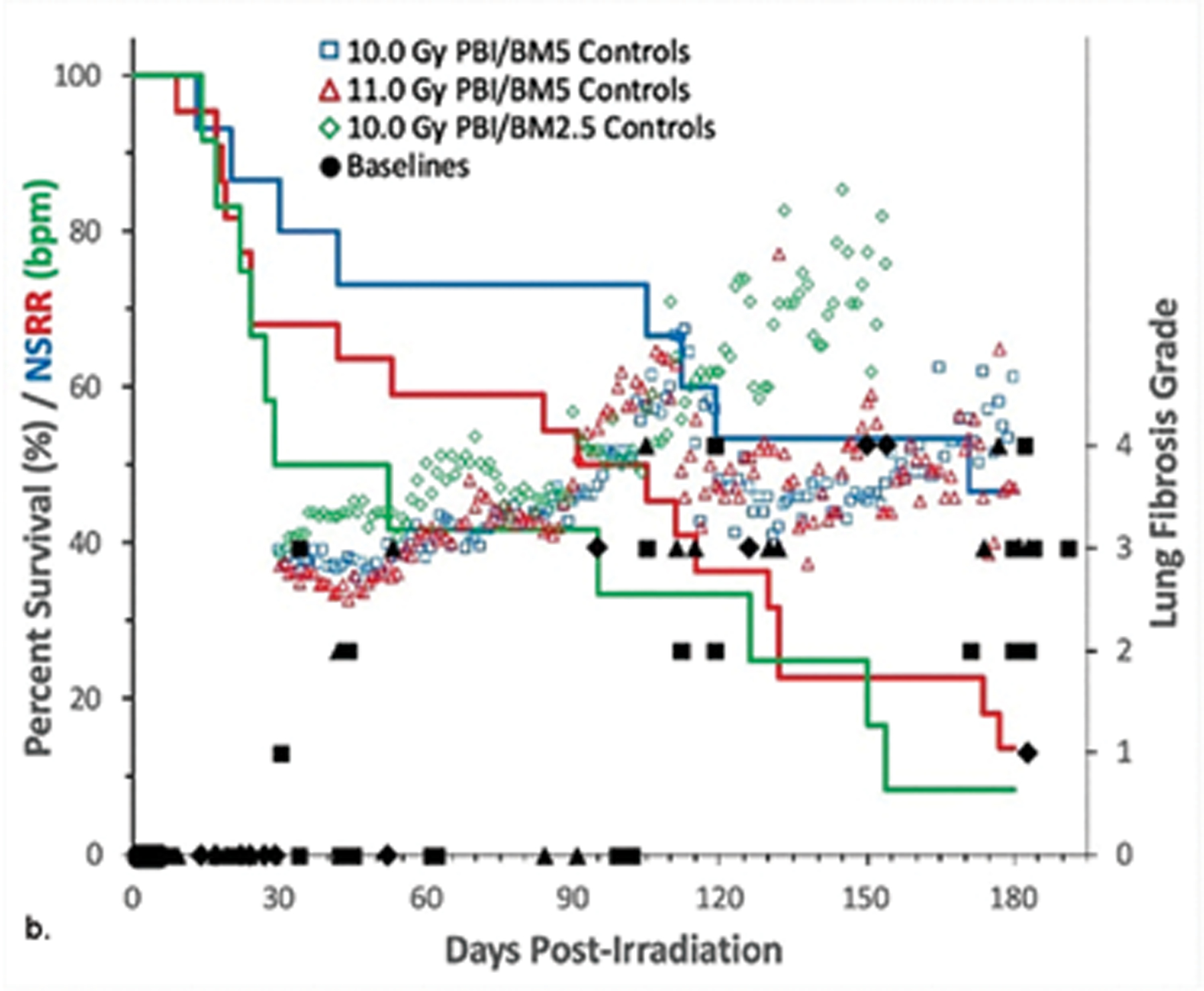
The nonhuman primate, rhesus macaque, is a relevant animal model that has been used to determine the efficacy of medical countermeasures to mitigate major signs of morbidity and mortality of radiation-induced lung injury. Herein, a literature review of published studies showing the evolution of lethal lung injury characteristic of the delayed effects of acute radiation exposure between the two significantly different exposure protocols, whole thorax lung irradiation and partial-body irradiation with bone marrow sparing in the nonhuman primate, is provided. The selection of published data was made from the open literature. The primary studies conducted at two research sites benefitted from the similarity of major variables; namely, both sites used rhesus macaques of approximate age and body weight and radiation exposure by LINAC-derived 6 MV photons at dose rates of 0.80 Gy min and 1.00 Gy min delivered to the midline tissue via bilateral, anterior/posterior, posterior/anterior geometry. An advantage relative to sex difference resulted from the use of male and female macaques by the Maryland and the Washington sites, respectively. Subject-based medical management was used for all macaques. The primary studies (6) provided adequate data to establish dose response relationships within 180 d for the radiation-induced lung injury consequent to whole thorax lung irradiation (male vs. female) and partial-body irradiation with bone marrow sparing exposure protocols (male). The dose response relationships established by probit analyses vs. linear dose relationships were characterized by two main parameters or dependent variables, a slope and LD50/180. Respective LD50/180 values for the primary studies that used whole thorax lung irradiation for respective male and female nonhuman primates were 10.24 Gy [9.87, 10.52] (n = 76, male) and 10.28 Gy [9.68, 10.92] (n = 40, female) at two different research sites. The respective slopes were steep at 1.73 [0.841, 2.604] and 1.15 [0.65, 1.65] probits per linear dose. The LD50/180 value and slope derived from the dose response relationships for the partial-body irradiation with bone marrow sparing exposure was 9.94 Gy [9.35, 10.29] (n = 87) and 1.21 [0.70, 1.73] probits per linear dose. A secondary study (1) provided data on limited control cohort of nonhuman primates exposed to whole thorax lung irradiation. The data supported the incidence of clinical, radiographic, and histological indices of the dose-dependent lung injury in the nonhuman primates. Tertiary studies (6) provided data derived from collaboration with the noted primary and secondary studies on control cohorts of nonhuman primates exposed to whole thorax lung irradiation and partial-body irradiation with bone marrow sparing exposure. These studies provided a summary of histological evidence of fibrosis, inflammation and reactive/proliferative changes in pneumonocytes characteristic of lung injury and data on biomarkers for radiation-induced lung injury based on matrix-assisted laser desorption ionization-mass spectrometry imaging and gene expression approaches. The available database in young rhesus macaques exposed to whole thorax lung irradiation or partial-body irradiation with bone marrow sparing using 6 MV LINAC-derived radiation with medical management showed that the dose response relationships were equivalent relative to the primary endpoint all-cause mortality. Additionally, the latency, incidence, severity, and progression of the clinical, radiographic, and histological indices of lung injury were comparable. However, the differences between the exposure protocols are remarkable relative to the demonstrated time course between the multiple organ injury of the acute radiation syndrome and that of the delayed effects of acute radiation exposure, respectively.


Conflict of interest statement

The authors declare no conflicts of interest.

Figures

Fig. 1.
Fig. 1.. Dose response relationship (DRR) for radiation-induced lung injury induced by partial-body irradiation with approximately 5% bone marrow sparing (PBI/BM5) or whole thorax lung irradiation (WTLI) exposure protocols in rhesus macaques.
UMSOM protocols. Rhesus macaques, male, were exposed to 6 MV LINAC-derived photons delivered to prescribed dose at midline tissue (xiphoid process) at a dose rate of 0.80 Gy min−1. SNBL protocols. Female macaques were exposed to 6 MV LINAC-derived photons delivered to prescribed dose at midline tissue at 1.0 ± 0.05 Gy min−1. All NHP received subject-based medical management as per respective IACUC-approved criteria and shared protocols. The DRRs were defined over time frames to assess organ-specific sub-syndromes over the 180 d study duration to assess radiation-induced lung injury characteristic of the DEARE. Irradiation protocols and exposures of WTLI (10.24 Gy) and PBI/BM5 (9.94 Gy) and WTLI (10.3 Gy) conducted at UMSOM and SNBL are compared.
Fig. 2.
Fig. 2.. Kaplan-Meier plot of survival probability demonstrating time-, dose- and protocol-dependent outcome relative to two exposure protocols in rhesus macaques.
Nonhuman primates at the UMSOM research site were exposed to uniform 6 MV LINAC-derived photons at a dose rate of 0.80 Gy min−1. All prescribed exposures were measured at midline tissue dose (xiphoid process). Animals received IACUC-approved, subject-based medical management to include dexamethasone. Two exposure protocols were used, PBI/BM5 and PBI/BM2.5, three doses of radiation and differential BM-sparing using PBI/BM5 10 Gy and 11 Gy or PBI/BM2.5 at 10 Gy or 12 Gy (PBI/BM2.5) or 12 Gy (PBI/BM5) respectively. A clear survival effect is noted between 10 and 11Gy using the PBI/BM5 protocol and between the PBI/BM5 and PBI/BM2.5 protocols at 10Gy.
Fig. 3.
Fig. 3.. Kaplan-Meier survival curves for rhesus macaque studies using the WTLI or PBI/BM2.5, PBI/BM5 exposure protocols: Relative all-cause mortality or survival probability over the 180 d study duration.
Nonhuman primates at the UMSOM research site were exposed to uniform 6 MV LINAC-derived photons at a dose rate of 0.80 Gy min−1. All prescribed exposures were measured at midline tissue dose (xiphoid process). Animals received IACUC-approved, subject-based medical management to include dexamethasone. Nonhuman primates were exposed to 10.5 Gy (n = 10), 10.74 Gy (n = 20) or 11 Gy (n = 10) using the WTLI protocol or 10 Gy (n = 15) or 11 Gy (n = 21) using the PBI/BM5 protocol or 10 Gy (n = 12) using the PBI/BM2.5 protocol at the UMSOM research site. Euthanasia was performed based on IACUC-defined criteria.
Fig. 4.
Fig. 4.. PBI/BM-sparing, the ARS-MOI time segment: The multi-organ ARS and clinical signs for NHP consequent to 10 Gy PBI/BM-sparing protocol.
Rhesus macaques were exposed to 10 Gy partial body irradiation with approximately 2.5% bone marrow sparing (PBI/BM2.5) by 6 MV LINAC-derived photons delivered to prescribed dose at midline tissue (xiphoid process) at a dose rate of 0.80 Gy min−1. Animals received IACUC-approved, subject-based medical management to include dexamethasone. The early clinical signs, to include dehydration, diarrhea, loss of body weight, cachexia, mucositis, plasma-based mediators and late occurring edema occur in the context of the overt multiple organ injury (MOI) characteristic of the acute radiation syndrome (ARS), e.g., GI-, H-ARS plus GI damage, immune suppression and acute kidney injury (AKI). The “early phase” clinical signs are presented in the context of the 180 d study duration. The time segment of the ARS represents the relatively “silent”, two mo latent period for MOI characteristic of the delayed effects of the acute radiation exposure (DEARE). The delayed edema and overt DEARE are evident at approximately 60 – 80 d post exposure (MacVittie et al. 2012; Farese et al. 2019).
Fig. 5.
Fig. 5.. The DEARE MOI time segment: The multi-organ injury within the DEARE and clinical signs for NHP consequent to 10 Gy – 11 Gy exposure using the WTLI protocol.
Rhesus macaques were exposed to 10 to 11 Gy whole thorax lung irradiation (WTLI), by 6 MV LINAC-derived photons delivered to prescribed dose at midline tissue (xiphoid process) at a dose rate of 0.80 Gy min−1. Animals received IACUC-approved, subject-based medical management to include dexamethasone. Lack of severe clinical signs noted in the “early phase” of the PBI/BM-sparing models is also evident in the context of the 180 d study duration after WTLI. The early time segment, 1 – 40 d, also represents the relatively “silent” or latent period for multiple organ injury (MOI) characteristic of the delayed effects of the acute radiation exposure (DEARE). The DEARE are characterized by lung-associated pleural effusion (PE), pneumonitis/fibrosis (PN/PF), (F) fibrosis and increased non-sedated respiratory rate (NSRR). The DEARE includes a prolonged and skewed repertoire of memory and naïve subsets of immune suppression since the thymus is within the WTLI field (Garofalo et al. 2014a; Garofalo et al. 2014b; MacVittie et al. 2014; MacVittie et al. 2017).
Fig. 6.
Fig. 6.. The ARS-DEARE time segment: The multi-organ injury within the ARS and DEARE and clinical signs for NHP consequent to 10 Gy PBI/BM2.5 exposure protocol.
Nonhuman primates at the UMSOM research site were exposed to 10 Gy partial-body irradiation with approximately 2.5% bone marrow sparing (PBI/BM2.5) by uniform 6 MV LINAC-derived photons at a dose rate of 0.80 Gy min−1. All prescribed exposures were measured at midline tissue dose (xiphoid process). Animals received IACUC-approved, subject-based medical management to include dexamethasone. The early clinical signs, to include dehydration, diarrhea, loss of body weight, cachexia, mucositis, plasma-based mediators and late occurring edema occur in the context of the overt multiple organ injury (MOI) characteristic of the acute radiation syndrome (ARS), e.g., GI-, H-ARS plus GI damage, immune suppression and acute kidney injury (AKI). The “early phase” clinical signs are presented in the context of the 180 d study duration. The early time segment also represents the relatively “silent”, two mo latent period for MOI characteristic of the delayed effects of the acute radiation exposure (DEARE). The DEARE are characterized by lung-associated pleural effusion (PE), pneumonitis/fibrosis (PF), fibrosis (F) and chronic kidney injury (CKI). The DEARE also include prolonged GI injury and a skewed repertoire of memory and naïve subsets of immune suppression. The delayed edema as overt DEARE was evident at approximately 60 – 80 d post exposure (MacVittie et al. 2014; Farese et al. 2019; MacVittie et al. 2019a).
Fig. 7.
Fig. 7.. Pulse oximetry, SpO2 values: Comparison, using a “best fit” line of mean SpO2 values over time post exposure for cohorts of NHP exposed to WTLI and PBI/BM-sparing protocols.
Rhesus macaques were exposed at 10 to 11 Gy, 6 MV LINAC-derived photons delivered to prescribed dose at midline tissue (xiphoid process) at a dose rate of 0.80 Gy min−1 to (a) whole thorax lung irradiation (WTLI) and (b) partial body irradiation with approximately 2.5% or 5% bone marrow sparing (PBI/BM). Animals received IACUC-approved, subject-based medical management to include dexamethasone. The oxygen saturation (SpO2) values are a measure of compensated respiratory function over the 180 d study duration (MacVittie et al. 2012; Garofalo et al. 2014a; MacVittie et al. 2017).
Fig. 8.
Fig. 8.. Kaplan-Meier survival curves for a) the PBI/BM-sparing exposure protocols and b) the WTLI exposure protocol: Relative mortality/survival probability and non-sedated respiratory rate (NSRR).
Rhesus macaques were exposed to (a) 10 to 11 Gy partial body irradiation (PBI) with approximately 2.5% or 5% bone marrow (BM) sparing, or (b) 10 to 11 Gy whole thorax lung irradiation (WTLI) by 6 MV LINAC-derived photons delivered to prescribed dose at midline tissue (xiphoid process) at a dose rate of 0.80 Gy min−1. Animals were observed through the 180d study duration. Animals received IACUC-approved, subject-based medical management to include dexamethasone. Changes in mean NSRR for each cohort are plotted as a function of time post exposure. This analysis was restricted to the NHP data sets that survived > 60 days post-exposure (e.g. survivors of GI-ARS and H-ARS coincident with prolonged GI) and for whom serial daily NSRR data were available. The mean time (d) to initiation of dexamethasone administration is color-coded relative to radiation dose and exposure protocol: 11 Gy PBI/BM5 (98), 10 Gy PBI/BM2.5 (98) and 10 Gy PBI/BM5 (127); WTLI at 10.74 Gy (78), 11 Gy (90), and 10 Gy (117d).
Fig. 9.
Fig. 9.. NSRR measured as breaths per minute in NHP exposed to PBI/BM-sparing and WTLI protocols.
Rhesus macaques were exposed at 10 to 11 Gy, 6 MV LINAC-derived photons delivered to prescribed dose at midline tissue (xiphoid process) at a dose rate of 0.80 Gy min−1 to either whole thorax lung irradiation (WTLI) or approximately 2.5% or 5% bone marrow sparing (PBI/BM). Animals received IACUC-approved, subject-based medical management to include dexamethasone. Changes in mean non-sedated respiratory rate (NSRR) are plotted as a function of time post irradiation. Cohorts are from contemporary sequential studies that included model development for PBI/BM5 and WTLI and those assessing the efficacy of a proprietary medical countermeasure. The analysis was restricted to NHPs that survive > 60 d post exposure. The mean first day of dexamethasone administration for each cohort are delineated by color-coded arrows: (a) WTLI 10 Gy (117 d), 10 Gy PBI/BM5 (127.7 d) and 10 Gy PBI/BM2.5 (98.4 d), and (b) 10.74 Gy WTLI (78 d), 11.0 Gy WTLI (90 d), and 11 Gy PBI/BM5 (98 d).
Fig. 10.
Fig. 10.. Radiographic analysis pneumonitis/fibrosis relative to total lung volume (PF:TLV) in NHP exposed to WTLI.
Rhesus macaques were exposed at 9 to 12 Gy whole thorax lung irradiation (WTLI), by 6 MV LINAC-derived photons delivered to prescribed dose at midline tissue (xiphoid process) at a dose rate of 0.80 Gy min−1. Radiographic analysis of lung injury by computed tomography (CT), showing (a) the mean ratio of volume of pneumonitis and fibrosis indexed against the total lung volume (PF:TLV) or (b) the mean ratio of volume of pleural effusion (PE) indexed against the total lung volume (PE:TLV) as a function of exposure dose and time post-exposure. CT scans were performed at baseline and every 30 days post-exposure until the end of study or until the animal was euthanized for cause. Subject-based dexamethasone administration and ongoing lethality of nonhuman primates (NHP) with greatest pulmonary injury influences the results present beyond d 30 (Garofalo et al. 2014a).
Fig. 11.
Fig. 11.. Comparative radiographic and clinical indices of pneumonitis/fibrosis relative to total lung volume (PF:TLV) and NSRR in NHP exposed to WTLI.
Rhesus macaques were exposed to 6 MV LINAC-derived photons delivered to prescribed dose at midline tissue (xiphoid process) at a dose rate of 0.80 Gy min−1 to (a) 10 to 11 Gy whole thorax lung irradiation (WTLI), or (b) 10 to 11 Gy partial body irradiation (PBI) with approximately 2.5% or 5% bone marrow (BM) sparing. Non-sedated respiratory rates (NSRR), based on the number of breaths per minute, were measured daily (scatter plots). Radiographic analysis of lung injury by computed tomography (CT), showing the mean ratio of volume of pneumonitis and fibrosis indexed against the total lung volume (PF:TLV) (solid lines) as a function of exposure dose and time post-exposure. CT scans were performed at baseline and every 30 d post-exposure until the end of study or until the animal met euthanasia criteria. Subject-based dexamethasone administration and ongoing lethality of nonhuman primates (NHP) with greatest pulmonary injury influences the results present beyond d 30 (Garofalo et al. 2014a; MacVittie et al. 2017).
Fig. 12.
Fig. 12.. Representative fields of lung tissue histology from NHP exposed to WTLI.
Representative lung tissue in a nonirradiated NHP showing normal lung morphology (A, C), and radiation-induced lung damage following 11.0 Gy whole thorax irradiation (WTLI) at 158 d post-exposure (b, d). Comparison of H&E (a, b) represent differences between a normal (a) and 11Gy irradiated (b) NHP. The irradiated NHP shows evidence of macrophage infiltration and congestion within lung architecture. Comparison of Masson’s Trichrome (c, d) stains demonstrates collagen deposition consistent with fibrosis in the 11.0 Gy irradiated NHP (d) as compared with the normal NHP (c). All images are shown at 20X magnification (Garofalo et al. 2014a).
Fig. 13.
Fig. 13.. Kaplan-Meier survival curves for the PBI/BM-sparing exposure protocol: Relative mortality/survival probability, histological time course of interstitial fibrosis and NSRR.
Rhesus macaques were exposed by 6 MV LINAC-derived photons delivered to prescribed dose at midline tissue (xiphoid process) at a dose rate of 0.80 Gy min−1 to (a) 10 or 11 Gy partial body irradiation with approximately 5% bone marrow sparing (PBI/BM5) or 10 Gy PBI/BM2.5. Animals received IACUC-approved, subject-based medical management to include dexamethasone. Lung sections were stained with Masson’s trichrome to identify deposition of collagen to assess latency, incidence and severity (grade). The mean values and grade of interstitial fibrosis as a function of time (d) post exposure [(a, b) solid symbols]. There was no noteworthy difference in the incidence of interstitial fibrosis in animals irradiated at 10 versus 11 Gy. Animals were euthanized for cause throughout the 180 d study duration. Non-sedated respiratory rates (NSRR), based on the number of breaths per minute, were measured daily [(b) open symbols]. This analysis was restricted to the NHP data set that survived > 60 d post-exposure (e.g. survivors of GI-ARS and H-ARS coincident with prolonged GI and for whom serial daily NSRR data were available) (MacVittie et al. 2012; Garofalo et al. 2014a).

Similar articles

Cited by

References

    1. Asano S. Multi-organ involvement: lessons from the experience of one victim of the Tokai-mura criticality accident. Br J Radiol 78; 2005.
    1. Baranov AE, Selidovkin GD, Butturini A, Gale RP. Hematopoietic recovery after 10-Gy acute total body radiation. Blood 83: 596–599; 1994. - PubMed
    1. Berkely FJ. Managing the adverse effects of radiation therapy. AM Fam Physician 82: 381–388; 2010. - PubMed
    1. Boittin F-X, Martigne P, Mayol J-F, Denis J, Raffin F, Coulon D, Grenier N, Drouet M, Herodin F. Experimental quantification of delayed radiation-indduced organ damage in highly irradiated rats with bone marrow protection: effect of radiation dose and organ sensitivity. Health Phys 109: 134–144; 2015. - PubMed
    1. Carter CL, Jones JW, Farese AM, MacVittie TJ, Kane MA. Lipidomic dysregulation within the lung parenchyma following whole-thorax lung irradiation: Markers of injury, inflammation and fibrosis detected by MALDI-MSI. Sci Rep 7: 10343; 2017. - PMC - PubMed

Publication types

MeSH terms