. 2020 Oct 2;11(1):4950.
doi: 10.1038/s41467-020-18750-9.

Remote ischemic conditioning counteracts the intestinal damage of necrotizing enterocolitis by improving intestinal microcirculation

Affiliations

Remote ischemic conditioning counteracts the intestinal damage of necrotizing enterocolitis by improving intestinal microcirculation

Yuhki Koike et al. Nat Commun. .

Abstract

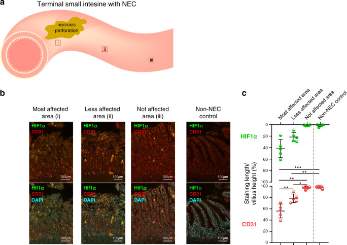
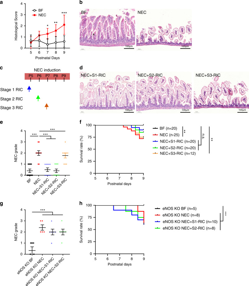
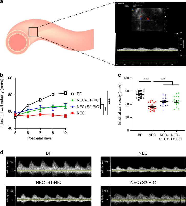
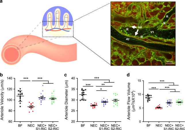
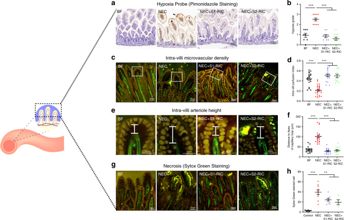
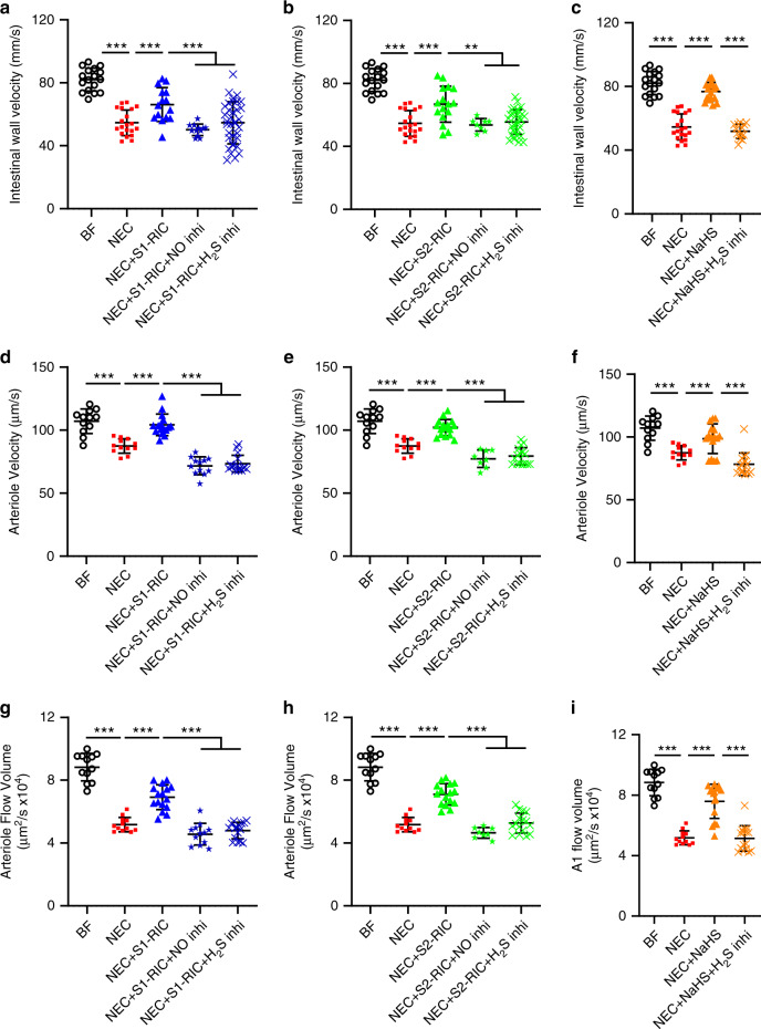
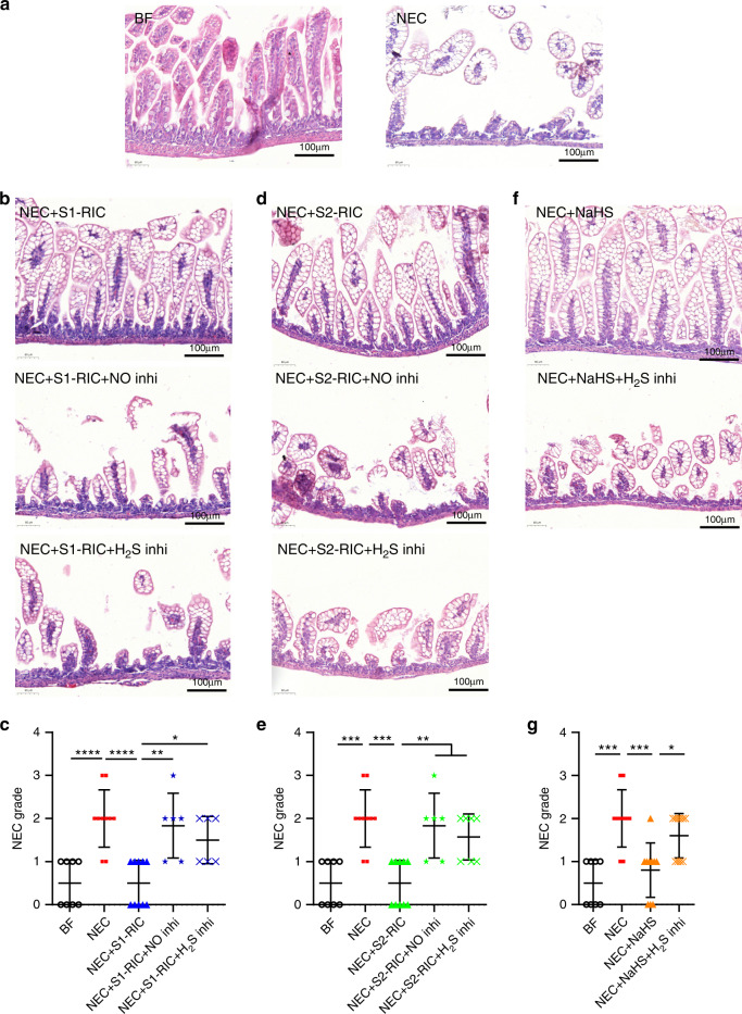
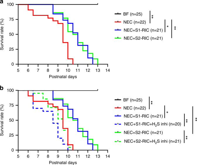
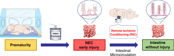
Necrotizing enterocolitis (NEC) is a devastating disease of premature infants with high mortality rate, indicating the need for precision treatment. NEC is characterized by intestinal inflammation and ischemia, as well derangements in intestinal microcirculation. Remote ischemic conditioning (RIC) has emerged as a promising tool in protecting distant organs against ischemia-induced damage. However, the effectiveness of RIC against NEC is unknown. To address this gap, we aimed to determine the efficacy and mechanism of action of RIC in experimental NEC. NEC was induced in mouse pups between postnatal day (P) 5 and 9. RIC was applied through intermittent occlusion of hind limb blood flow. RIC, when administered in the early stages of disease progression, decreases intestinal injury and prolongs survival. The mechanism of action of RIC involves increasing intestinal perfusion through vasodilation mediated by nitric oxide and hydrogen sulfide. RIC is a viable and non-invasive treatment strategy for NEC.


Conflict of interest statement

The authors declare no competing interests.

Figures

Fig. 1
Fig. 1. Human NEC is associated with mucosal hypoxia and impairment of the microvasculature.
a The terminal ileum of NEC patients (n = 5) and non-NEC control patients (n = 5) were analyzed and compared using immunofluorescence staining. b, c The intestine was divided into three regions: (i) the most affected ileum close to the area of necrosis and perforation (n = 5), (ii) the less affected ileum distant from the necrosis/perforation (n = 5), and (iii) the not affected ileum farther away from the damaged area (n = 5). The most affected ileum had the highest expression of hypoxia-inducible factor 1α (HIF1α) and the lowest expression of endothelial cell marker (CD31), compared to less affected, not affected areas and non-NEC controls. There was a progressive decrease in HIF1α expression and progressive increase in CD31 expression from NEC-damaged to less damaged area. Immunofluorescence staining and imaging and analysis of slides were performed by blinded investigators and data werr compared using two-sided one-way ANOVA with post hoc Turkey test and repeated measure for the comparison of different areas within the NEC patient and no pairing comparison to the non-NEC control (*p < 0.05, **p < 0.01, ***p < 0.001). Scale bars are equivalent to 100 µm in all the images shown, and data are presented as mean ± SEM. Experiments were repeated independently 3 times, with similar results. Source data are provided as a Source Data file.
Fig. 2
Fig. 2. RIC improves intestinal damage during experimental NEC.
a To establish the timing of RIC regimen in the initial development of NEC, intestinal injury was investigated in C57BL/6 mouse pups from P5 to P9 (BF P5–P8: n = 6; BF P9: n = 10; NEC P5: n = 6; NEC P6–8: n = 7; NEC P9: n = 10). b Morphology of the ileum was compared between breastfed (BF) control (n = 10) and NEC pups (n = 10) using hematoxylin and eosin staining. Scale bars are equivalent to 100 µm in the images shown. Experiments were repeated independently 3 times, with similar results. c RIC was given in three distinct time points, each consisting of two episodes of RIC that are 48 h apart: RIC was initiated on P5 (Stage 1, n = 10), P6 (Stage 2, n = 10), or P7 (Stage 3 RIC, n = 10). d Effect of Stages 1–3 RIC on intestinal morphology were compared in NEC pups at P9. Scale bars are equivalent to 100 µm in the images shown. e Histological slides were graded for BF (n = 8), NEC (n = 10), NEC + Stage 1 RIC (n = 10), NEC + Stage 2 RIC (n = 10), and NEC + Stage 3 RIC (n = 10) by 3 investigators blinded to treatment allocation based on the NEC histopathological scoring system. Mice with histological grade ≥2 were considered to have NEC. f Stages 1 and 2 RIC equally enhanced survival of NEC pups up to sacrifice on P9. Stage 3 RIC did not improve NEC survival. g Effect of Stages 1 and 2 RIC on intestinal morphology in eNOS knockout NEC pups (eNOS BF: n = 5, eNOS NEC: n = 5, eNOS NEC + Stage 1 RIC: n = 6, eNOS NEC + Stage 2 RIC: n = 6). Histological slides were graded by 3 blinded investigators as described above. h eNOS knockout pups with NEC receiving Stage 1 or 2 RIC had a similar survival rate to eNOS knockout pups with NEC alone. Data were compared using two-sided one-way ANOVA with post hoc Turkey test (*p < 0.05; **p < 0.01, ***p < 0.001). Data are presented as mean ± SEM. Source data are provided as a Source Data file.
Fig. 3
Fig. 3. RIC improves intestinal wall perfusion during NEC development.
a Intestinal wall perfusion was measured using Doppler ultrasound and calculated as average flow velocity (mm/s) of multiple abdominal regions. b Intestinal wall perfusion measured daily from P5 to P9 showed reduced perfusion in NEC pups which was improved following conditioning with Stage 1 or 2 RIC (n = 6 per group; minimum of 3 readings of different abdominal quadrants were obtained per pup; **p < 0.01; ***p < 0.001). c Intestinal wall perfusion measured on P9 in NEC pups showed reduced perfusion compared to breastfed (BF) control, which was preserved in NEC pups receiving Stage 1 or 2 RIC (n = 6 per group; minimum of 2 readings of different abdominal quadrants were obtained per pup; **p < 0.01; ***p < 0.001). d Representative images of arterial waves obtained with the Doppler ultrasound in P9 pups from listed groups. Data were compared using two-sided one-way ANOVA with post hoc Turkey test and data are presented as mean ± SEM. Source data are provided as a Source Data file.
Fig. 4
Fig. 4. RIC preserves intestinal microcirculation in the submucosa.
a Depiction of submucosal arterioles (A1) and submucosal venules (V1) recorded via in vivo live imaging by TPLSM. Experiments were repeated independently 3 times, with similar results. Arteriole response in the ileal submucosa at P9 in terms of arteriole b blood flow velocity (µm/s), c diameter (µm), and d flow volume [(μm)3/s] compared between breastfed (BF) control, NEC, NEC with Stage 1 RIC, and NEC with Stage 2 RIC groups (BF and NEC: n = 6; NEC + Stage 1 or 2 RIC: n = 8; minimum of 2 readings were obtained per group). Data were compared using two-sided one-way ANOVA with post hoc Turkey test (*p < 0.05; **p < 0.01; ****p < 0.0001). Data are presented as mean ± SEM. Source data are provided as a Source Data file.
Fig. 5
Fig. 5. RIC improves intestinal villi microvasculature and reduces ischemia and necrosis of enterocytes at the villi tip.
a, b Pups were injected with pimonidazole, a sensitive marker which allows localization of intestinal ischemia. Data represent immunohistochemistry of pimonidazole in the ileum comparing breastfed (BF) control (n = 8), NEC (n = 10), NEC with Stage 1 RIC (n = 10), and NEC with Stage 2 RIC (n = 10). Scale bars are equivalent to 100 µm in the images shown. c Intra-villi microvasculature was investigated in RosamT/mG/+;Tie2-Cre using TPLSM and compared between BF, NEC, NEC with Stage 1 RIC, and NEC with Stage 2 RIC (n = 4 per group; and minimum of 2 measurements were obtained per group). d Villus perfusion index was calculated as the ratio of the area of intra-villi arterioles to the area of the whole villi and compared between the listed groups. e, f The distance between the apex of the capillary loop and the apical side of the villi epithelium was measured and compared between listed groups. g Cell death (yellow staining in the tip of villi) in the ileal epithelium was detected using Sytox Green, a marker of necrosis which does not permeate into live cells but binds to cellular nucleic acids in dead cells, staining them with intense green fluorescence and was compared between listed groups. h Necrotic cells were counted in the listed groups (*p < 0.05; **p < 0.01; ****p < 0.0001). Data were compared using two-sided one-way ANOVA with post hoc Turkey test (**p < 0.01; ****p < 0.0001). Scale bars in (c, e and g are equivalent to 50 µm, and data are presented as mean ± SEM. Source data are provided as a Source Data file.
Fig. 6
Fig. 6. RIC preserves intestinal microcirculation via endogenous vasodilators nitric oxide and hydrogen sulfide.
Administration of NO-synthase inhibitor and H2S-synthesizing enzyme inhibitors abolished a Stage 1 RIC and b Stage 2 RIC-mediated preservation of intestinal wall flow velocity (mm/s) measured with Doppler ultrasound. c Treatment with NaHS, exogenous donor of H2S, improved intestinal wall flow velocity (mm/s), but not in the presence of H2S-synthesizing enzyme inhibitors. Administration of NO-synthase inhibitor and H2S-synthesizing enzyme inhibitors abolished the d, g Stage 1 RIC- and e, h Stage 2 RIC-mediated preservation of submucosal arteriole velocity (µm/s) and arteriole flow volume [(μm)3/s] respectively, measured using TPLSM in real time. f, i Treatment with NaHS improved submucosal arteriole velocity (µm/s) and arteriole flow volume [(μm)3/s] respectively, but not following administration of H2S-synthesizing enzyme inhibitors. Data were compared using two-sided one-way ANOVA with post hoc Turkey test (n = 8 per group; minimum of 2 readings were obtained per group; **p < 0.01; ***p < 0.001). Data are presented as mean ± SEM. Source data are provided as a Source Data file.
Fig. 7
Fig. 7. RIC-mediated preservation of intestinal perfusion via nitric oxide and hydrogen sulfide is required to improve intestinal injury during NEC.
a Morphology of the ileum was assessed using hematoxylin and eosin staining in breastfed (BF) (n = 8) control and NEC pups (n = 10). Intestinal injury increased in NEC pups receiving b, c Stage 1 RIC following administration of inhibitors of NO-synthase (n = 6) or H2S-synthesizing enzymes (n = 6), as well as in pups receiving d, e Stage 2 RIC following administration of inhibitors of NO-synthase (n = 6) or H2S-synthesizing enzymes (n = 7) compared to NEC pups receiving Stage 1 RIC (n = 10) or Stage 2 RIC (n = 10) without drug treatment. Morphology of the ileum was assessed using hematoxylin and eosin staining and histological slides were graded by 3 investigators blinded to treatment allocation based on the NEC histopathological scoring system that defines mice with NEC grade ≥ 2 as NEC positive. f, g Treatment with NaHS improved intestinal injury in NEC pups, resulting in a lower NEC grade, but not following treatment with H2S-synthesizing enzyme inhibitors (BF: n = 8; NEC: n = 10; NEC + NaHS: n = 10, NEC + NaHS+H2S-synthesizing enzyme inhibitors: n = 10). Data were compared using two-sided one-way ANOVA with post hoc Turkey test (*p < 0.05; **p < 0.01; ***p < 0.001). Scale bars are equivalent to 100 µm in a, b, d and f. Data are presented as mean ± SEM. Source data are provided as a Source Data file.
Fig. 8
Fig. 8. RIC enhanced survival during and after NEC and this effect was abolished following administration of H2S-synthesizing enzyme inhibitors.
a After P9, pups from all groups except breastfed (BF) controls, continued to receive gavage feeding three times per day by single investigator blinded to treatment allocation and were monitored until death occurred. Pups receiving Stage 1 or 2 RIC survived significantly longer after NEC induction (*p < 0.05; **p < 0.01). b Survival rate after P9 was similar between NEC pups and NEC pups receiving Stage 1 or 2 RIC and given H2S-synthesizing enzyme inhibitors (*p < 0.05; **p < 0.01). Survival curves were compared using the logrank test (*p < 0.05; **p < 0.01). Data are presented as mean ± SEM. Source data are provided as a Source Data file.
Fig. 9
Fig. 9. Remote ischemic conditioning counteracts the intestinal damage of necrotizing enterocolitis (NEC) by improving intestinal microcirculation.
Prematurity and formula feeding are among the main risk factors contributing to the development of NEC, a devastating disease of premature infants characterized by intestinal inflammation and ischemia. NEC is characterized by derangements in intestinal microcirculatory blood flow, villus core separation, sloughing of the villi, and presence of necrotic tissue at the villi. Remote ischemic conditioning is a therapeutic maneuver whereby application of brief cycles of ischemia and reperfusion to a limb protects a distant organ from sustained ischemic damage. During the initial stage of experimental NEC, remote ischemic conditioning improves intestinal injury, reduces inflammation, and enhances survival. The mechanism of action of remote ischemic conditioning involves preservation of intestinal microcirculation that is mediated by the vasodilators, hydrogen sulfide and nitric oxide. Remote ischemic conditioning is a noninvasive treatment strategy for neonatal NEC.

Similar articles

Cited by

References

    1. Alganabi, M., Lee, C., Bindi, E., Li, B. & Pierro, A. Recent advances in understanding necrotizing enterocolitis. F1000Res8, 10.12688/f1000research.17228.1 (2019). - PMC - PubMed
    1. Neu J, Walker WA. Necrotizing enterocolitis. N. Engl. J. Med. 2011;364:255–264. doi: 10.1056/NEJMra1005408. - DOI - PMC - PubMed
    1. Rees CM, Pierro A, Eaton S. Neurodevelopmental outcomes of neonates with medically and surgically treated necrotizing enterocolitis. Arch. Dis. Child Fetal Neonatal Ed. 2007;92:F193–F198. doi: 10.1136/adc.2006.099929. - DOI - PMC - PubMed
    1. Nino, D. F. et al. Cognitive impairments induced by necrotizing enterocolitis can be prevented by inhibiting microglial activation in mouse brain. Sci. Transl. Med.10, 10.1126/scitranslmed.aan0237 (2018). - PMC - PubMed
    1. Squires RH, et al. Natural history of pediatric intestinal failure: initial report from the Pediatric Intestinal Failure Consortium. J. Pediatr. 2012;161:723–728 e722. doi: 10.1016/j.jpeds.2012.03.062. - DOI - PMC - PubMed

Publication types