

Development of an electronic navigation system for elimination of examiner-dependent factors in the ultrasound screening for developmental dysplasia of the hip in newborns
- PMID: 33009470
- PMCID: PMC7532432
- DOI: 10.1038/s41598-020-73536-9
Development of an electronic navigation system for elimination of examiner-dependent factors in the ultrasound screening for developmental dysplasia of the hip in newborns
Abstract
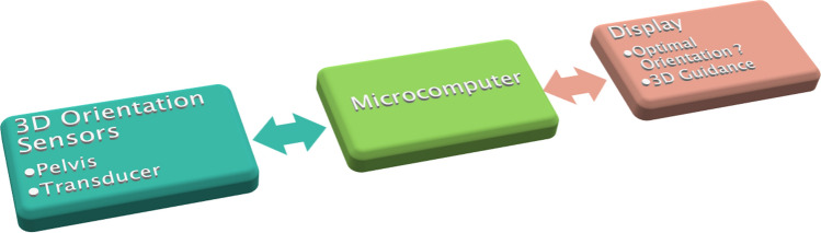
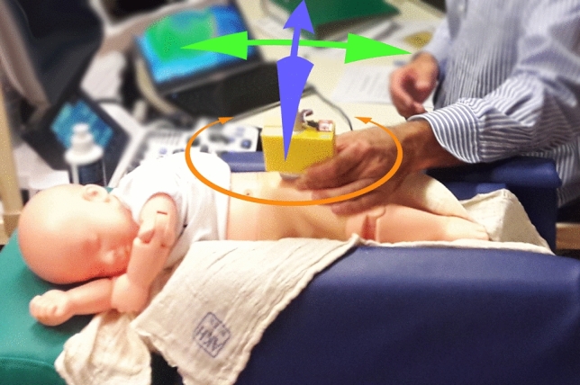
To develop an electronic navigation system to increase reliability and comparability in the ultrasound screening of developmental dysplasia of the hip (DDH). The impact of the navigation system on transducer positioning and on sonographic measurements according to Graf was analyzed. Twenty hips in newborns were examined sonographically using a new navigation system capable of detecting the transducer and pelvis position in order to calculate the relative tilt in the frontal, axial, and sagittal-plane. In each newborn an ultrasound image was obtained conventionally according to Graf and a second image using the sonographic navigation system. Relative roll and pitch angles and sonographic measurements were analyzed using paired T-tests and Levene-tests. Relative tilt angles in the conventional group ranged from - 8.9° to 14.3° (frontal-plane) and - 23.8° to 14.2° (axial-plane). In the navigation-assisted group ranges from - 3.0° to 3.5° and - 2.8° to 4.5° were observed. Variances were significantly lower in the navigation-assisted group (p < 0.001 and p = 0.004 respectively). The navigation system allowed for a significant reduction of relative tilt angles between the transducer and the newborn pelvis, thus supporting an optimal transducer positioning. This is a promising approach to improve reproducibility and reliability in the ultrasound screening for DDH.
Conflict of interest statement
On behalf of all authors, the corresponding author states that there is no conflict of interest. One of the authors (RW) has received institutional funding from Stryker, DePuy Synthes Austria, Johnson & Johnson Medical Limited, Medacta, Zimmer Biomet Gmbh.
Figures


References
-
- Simon EA, et al. Inter-observer agreement of ultrasonographic measurement of alpha and beta angles and the final type classification based on the Graf method. Swiss Med. Wkly. 2004;134:671–677. - PubMed
-
- Roposch A, Graf R, Wright JG. Determining the reliability of the Graf classification for hip dysplasia. Clin. Orthop. Relat. Res. 2006;447:119–124. doi: 10.1097/01.blo.0000203475.73678.be. - DOI - PubMed
Publication types
MeSH terms
LinkOut - more resources
Full Text Sources
Research Materials
