. 2020 Nov;24(22):13289-13302.
doi: 10.1111/jcmm.15946. Epub 2020 Oct 3.

Exosomes derived from smooth muscle cells ameliorate diabetes-induced erectile dysfunction by inhibiting fibrosis and modulating the NO/cGMP pathway

Affiliations

Exosomes derived from smooth muscle cells ameliorate diabetes-induced erectile dysfunction by inhibiting fibrosis and modulating the NO/cGMP pathway

Jingyu Song et al. J Cell Mol Med. 2020 Nov.

Abstract

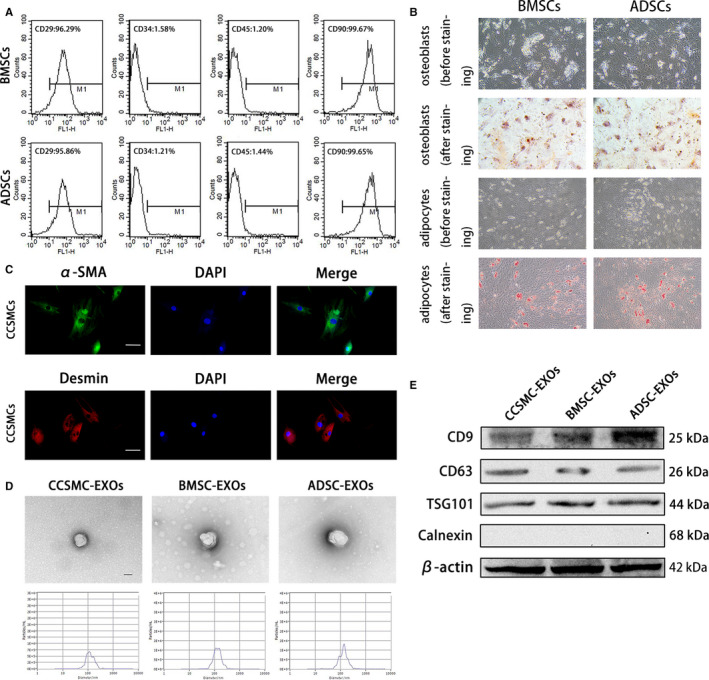
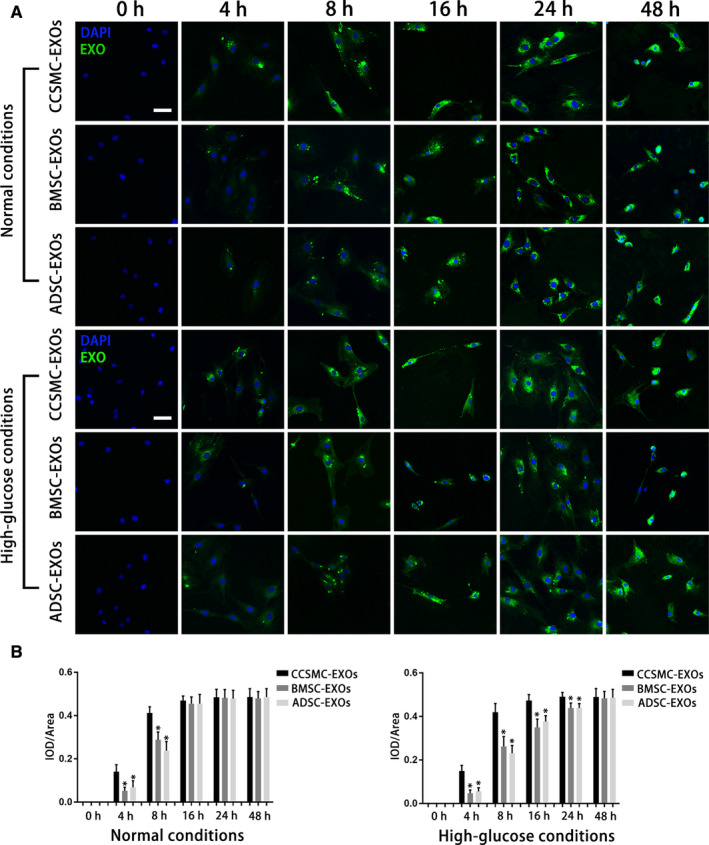
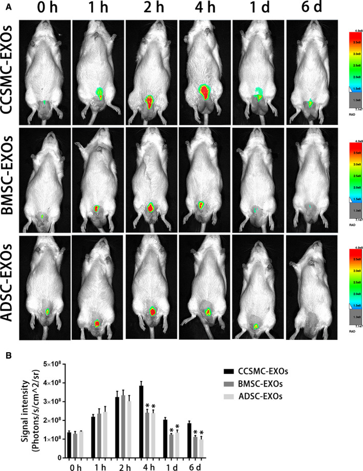
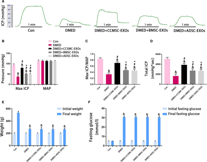
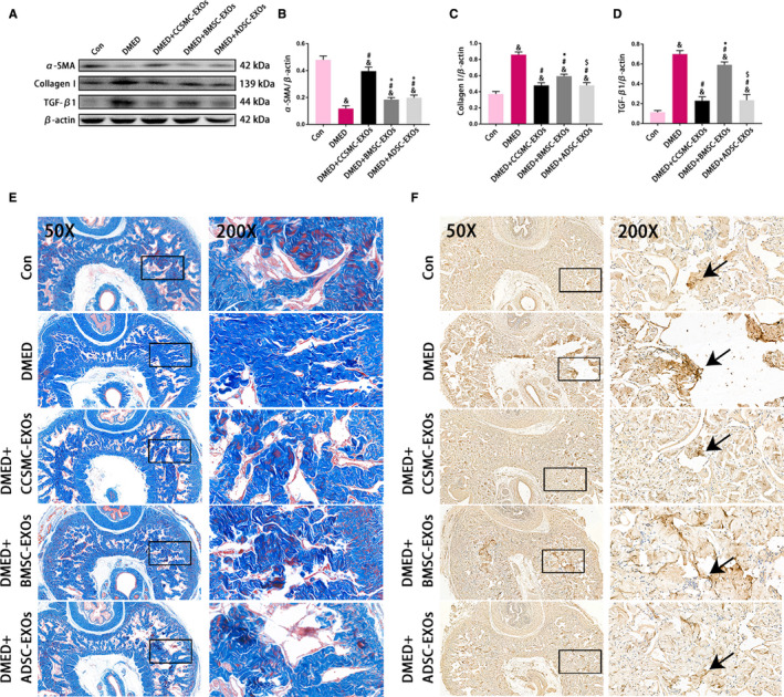
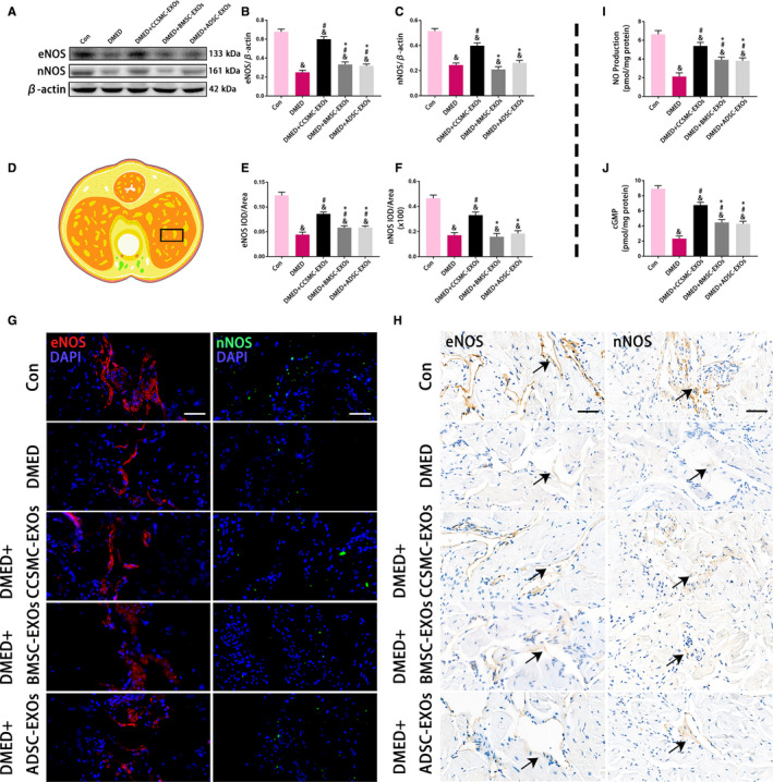
Erectile dysfunction (ED) is a major health issue among men with diabetes, and ED induced by diabetes mellitus (DMED) is particularly difficult to treat. Therefore, novel therapeutic approaches for the treatment of DMED are urgently needed. Exosomes, nanosized particles involved in many physiological and pathological processes, may become a promising tool for DMED treatment. In this study, we investigated the therapeutic effect of exosomes derived from corpus cavernosum smooth muscle cells (CCSMC-EXOs) on erectile function in a rat model of diabetes and compared their effect with that of exosomes derived from mesenchymal stem cells (MSC-EXOs). We incubated labelled CCSMC-EXOs and MSC-EXOs with CCSMCs and then observed uptake of the exosomes at different time points using laser confocal microscopy. CCSMC-EXOs were more easily taken up by CCSMCs. The peak concentration and retention time of labelled CCSMC-EXOs and MSC-EXOs in the corpus cavernosum of DMED rats after intracavernous injection were compared by in vivo imaging techniques. Intracavernous injection of CCSMC-EXOs was associated with a relatively high peak concentration and long retention time. Our data showed that CCSMC-EXOs could improve erectile function in DMED rats. Meanwhile, CCSMC-EXOs could exert antifibrotic effects by increasing the smooth muscle content and reducing collagen deposition. CCSMC-EXOs also increased the expression of eNOS and nNOS, followed by increased levels of NO and cGMP. These findings initially identify the possible role of CCSMC-EXOs in ameliorating DMED through inhibiting corporal fibrosis and modulating the NO/cGMP signalling pathway, providing a theoretical basis for a breakthrough in the treatment of DMED.

Keywords: NO/cGMP signalling pathway; diabetes; erectile dysfunction; exosomes; fibrosis.


Conflict of interest statement

The authors confirm that there are no conflict of interest.

Figures

FIGURE 1
FIGURE 1
Cell identification and exosome characterization. (A) Representative flow cytometry histograms of BMSCs and ADSCs show positive staining for CD29 and CD90 but not for CD34 and CD45. (B) BMSCs and ADSCs were successfully induced into osteoblasts (positively stained with Alizarin Red S) and adipocytes (positively stained with Oil Red O). The magnification is 100×. (C) Representative immunofluorescence results of CCSMCs show positive expression for α‐SMA and desmin. Scale bars = 50 μm. (D) Exosomes derived from CCSMCs, BMSCs and ADSCs were observed using transmission electron microscopy, and the particle size distributions of the exosomes were measured by nanoparticle tracking analysis. Scale bars = 100 nm. (E) Representative results of Western blot analysis of exosomes derived from CCSMCs, BMSCs and ADSCs show positive expression for CD9, CD63 and TSG101 but not for calnexin. CCSMC: corpus cavernosum smooth muscle cell; BMSC: bone marrow stem cell; ADSC: adipose‐derived stem cell; CCSMC‐EXOs: exosomes derived from corpus cavernosum smooth muscle cells; BMSC‐EXOs: exosomes derived from bone marrow stem cells; ADSC‐EXOs: exosomes derived from adipose‐derived stem cells; α‐SMA: α‐smooth muscle actin; DAPI: 4’,6‐diamidino‐2‐phenylindole
FIGURE 2
FIGURE 2
Uptake of exosomes by CCSMCs. (A) Representative confocal images of PKH67‐labelled exosomes taken up by CCSMCs cultured under different conditions are shown. The culture time is indicated. Scale bars = 50 μm. (B) Time courses of the uptake of different types of exosomes by CCSMCs under different conditions are presented as bar graphs. Data are expressed as the mean ± standard deviation. * P < .05 compared with the CCSMC‐EXO group. CCSMC‐EXOs: exosomes derived from corpus cavernosum smooth muscle cells; BMSCs‐EXOs: exosomes derived from bone marrow stem cells; ADSC‐EXOs: exosomes derived from adipose‐derived stem cells; DAPI: 4',6‐diamidino‐2‐phenylindole; IOD: integral optical density; IOD/area: mean optical density
FIGURE 3
FIGURE 3
Exosome intracavernous injection and in vivo imaging. (A) Representative images of DMED rats that underwent intracavernous injection with DiR‐labelled exosomes. (B) The signal intensities of different DiR‐labelled exosomes in DMED rats at different time points after injection are presented as a bar graph. Data are expressed as the mean ± standard deviation. * P < .05 compared with the CCSMC‐EXO group. CCSMC‐EXOs: exosomes derived from corpus cavernosum smooth muscle cells; BMSC‐EXOs: exosomes derived from bone marrow stem cells; ADSC‐EXOs: exosomes derived from adipose‐derived stem cells; DMED: diabetes mellitus‐induced erectile dysfunction
FIGURE 4
FIGURE 4
Evaluation of erectile function and metabolic indexes. (A) Representative ICP traces measured by stimulating the cavernous nerves with 5 V for 1 min. (B) The max ICP and MAP values for each group are presented as a bar graph. (C) The max ICP/MAP values for each group are presented as a bar graph to show the erectile function. (D) The total ICP values for each group are presented as a bar graph to show the erectile function. (E) Initial and final weights for each group are presented as a bar graph. (F) Initial and final fasting glucose levels for each group are presented as a bar graph. Data are expressed as the mean ± standard deviation. & P < .05 compared with the Con group. # P < .05 compared with the DMED group. * P < .05 compared with the DMED + CCSMC‐EXO group. CCSMC‐EXOs: exosomes derived from corpus cavernosum smooth muscle cells; BMSC‐EXOs: exosomes derived from bone marrow stem cells; ADSC‐EXOs: exosomes derived from adipose‐derived stem cells; DMED: diabetes mellitus‐induced erectile dysfunction; ICP: intracavernous pressure; MAP: mean arterial pressure
FIGURE 5
FIGURE 5
Evaluation of corporal fibrosis in the corpus cavernosum. (A) Representative results of Western blot analysis of α‐SMA, Collagen I and TGF‐β1 in rats from all five groups. (B) Expression of α‐SMA in all five groups with β‐actin as the loading control is presented as a bar graph. (C) Expression of Collagen I in all five groups with β‐actin as the loading control is presented as a bar graph. (D) Expression of TGF‐β1 in all five groups with β‐actin as the loading control is presented as a bar graph. (E) Representative images showing smooth muscle and collagen staining in all five groups. Smooth muscle and collagen in the corpus cavernosum are stained red and blue, respectively. Original magnification, 50× and 200×. (F) Representative results of immunohistochemistry for TGF‐β1 in all five groups (50× and 200×). The arrows indicate TGF‐β1 expression. Data are expressed as the mean ± standard deviation. & P < .05 compared with the Con group. # P < .05 compared with the DMED group. * P < .05 compared with the DMED + CCSMC‐EXO group. $ P < .05 compared with the DMED + BMSC‐EXO group. CCSMC‐EXOs: exosomes derived from corpus cavernosum smooth muscle cells; BMSC‐EXOs: exosomes derived from bone marrow stem cells; ADSC‐EXOs: exosomes derived from adipose‐derived stem cells; DMED: diabetes mellitus‐induced erectile dysfunction
FIGURE 6
FIGURE 6
eNOS and nNOS expression with the NO and cGMP concentration in the corpus cavernosum. (A) Representative results of Western blot analysis of eNOS and nNOS in the rats of all five groups. (B) Expression of eNOS in all five groups with β‐actin as the loading control is presented as a bar graph. (C) Expression of nNOS in all five groups with β‐actin as the loading control is presented as a bar graph. (D) The black rectangle indicates the area of the corpus cavernosum selected for comparison. (E) The eNOS level is defined as eNOS IOD/area according to the immunofluorescence results. (F) The nNOS level is defined as nNOS IOD/area according to the immunofluorescence results. (G) Representative results of immunofluorescence analysis of eNOS and nNOS in all five groups. Scale bars = 50 μm. (H) Representative results of immunohistochemistry analysis of eNOS and nNOS in all five groups. The arrows indicate eNOS and nNOS expression (Because the expression levels of eNOS and nNOS in DMED group were very low, the coloring was very light). Scale bars = 50 μm. (I) The NO production in all five groups was determined with an assay kit. (J) The cGMP concentration in all five groups was determined with an assay kit. Data are expressed as the mean ± standard deviation. & P < .05 compared with the Con group. # P < .05 compared with the DMED group. * P < .05 compared with the DMED + CCSMC‐EXO group. CCSMC‐EXOs: exosomes derived from corpus cavernosum smooth muscle cells; BMSC‐EXOs: exosomes derived from bone marrow stem cells; ADSC‐EXOs: exosomes derived from adipose‐derived stem cells; DMED: diabetes mellitus‐induced erectile dysfunction; DAPI: 4',6‐diamidino‐2‐phenylindole; eNOS: endothelial nitric oxide synthase; nNOS: neuronal nitric oxide synthase; NO: nitric oxide; cGMP: cyclic guanosine monophosphate; IOD: integral optical density; IOD/area: mean optical density

References

    1. Thorve VS, Kshirsagar AD, Vyawahare NS, Joshi VS, Ingale KG, Mohite RJ. Diabetes‐induced erectile dysfunction: epidemiology, pathophysiology and management. J Diabetes Complications. 2011;25:129‐136. - PubMed
    1. Simeni Njonnou SR, Boombhi J, Etoa Etoga MC, et al. Prevalence of diabetes and associated risk factors among a group of prisoners in the Yaoundé central prison. J Diabetes Res. 2020;2020:5016327. - PMC - PubMed
    1. Shamloul R, Ghanem H. Erectile dysfunction. Lancet. 2013;381:153‐165. - PubMed
    1. Malavige LS, Levy JC. Erectile dysfunction in diabetes mellitus. J Sex Med. 2009;6:1232‐1247. - PubMed
    1. Cui K, Tang Z, Li C‐C, et al. Lipoxin A4 improves erectile dysfunction in rats with type I diabetes by inhibiting oxidative stress and corporal fibrosis. Asian J Androl. 2018;20:166‐172. - PMC - PubMed

Publication types