

Short-term outcomes of patients with neovascular exudative AMD: the effect of COVID-19 pandemic
- PMID: 33009973
- PMCID: PMC7532341
- DOI: 10.1007/s00417-020-04955-7
Short-term outcomes of patients with neovascular exudative AMD: the effect of COVID-19 pandemic
Abstract
Purpose: To estimate the impact of delayed care during the coronavirus disease 2019 (COVID-19) pandemic on the outcomes of patients with neovascular age-related macular degeneration (AMD).
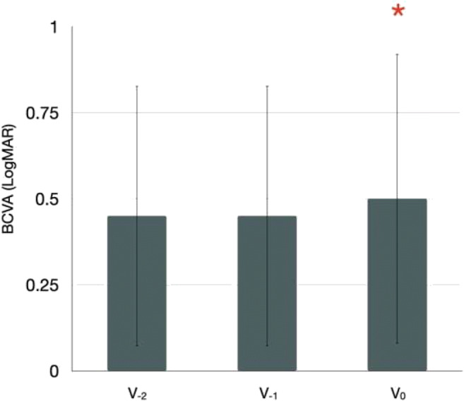
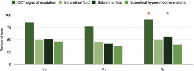
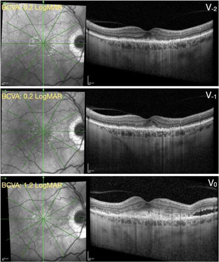
Methods: Consecutive patients with diagnosis of neovascular AMD were consecutively enrolled between March 9, 2020, and June 12, 2020, (during and immediately after the Italian COVID-19 quarantine). During the inclusion (or pandemic) visit (V0), patients received a complete ophthalmologic evaluation, including optical coherence tomography (OCT). Best-corrected visual acuity (BCVA) and OCT findings from the two preceding visits (V-1 and V-2) were compared with data at V0.
Results: One-hundred patients (112 eyes) were enrolled in this study. The time interval between following visits was 110.7 ± 37.5 days within V0 and V-1 and 80.8 ± 39.7 days within V-1 and V-2, respectively (P < 0.0001). BCVA was statistically worse at the V0 visit as compared with the immediately preceding (V-1) visit (0.50 ± 0.43 LogMAR and 0.45 ± 0.38 LogMAR at the V0 and V-1 visits, respectively; P = 0.046). On structural OCT, 91 out of 112 (81.2%) neovascular AMD eyes displayed the evidence of exudative disease activity at the V0 visit, while 77 (68.7%) eyes exhibited signs of exudation at the V-1 visit (P = 0.022). No differences in terms of BCVA and OCT findings were detected between the V-1 and V-2 visits. In multiple regression analysis, the difference in BCVA between V0 and V-1 visits was significantly associated with the interval time within these two visits (P = 0.026).
Conclusion: The COVID-19 pandemic-related postponement in patient care proved to be significantly associated with worse short-term outcomes in these patients.
Keywords: COVID-19; Neovascular AMD; Outcome; Retina.
Conflict of interest statement
Francesco Bandello is a consultant for Alcon (Fort Worth, Texas, USA), Alimera Sciences (Alpharetta, Georgia, USA), Allergan Inc. (Irvine, California, USA), Farmila-Thea (Clermont-Ferrand, France), Bayer Shering-Pharma (Berlin, Germany), Bausch And Lomb (Rochester, New York, USA), Genentech (San Francisco, California, USA), Hoffmann-La-Roche (Basel, Switzerland), Novagali Pharma (Évry, France), Novartis (Basel, Switzerland), Sanofi-Aventis (Paris, France), Thrombogenics (Heverlee, Belgium), Zeiss (Dublin, USA). Giuseppe Querques is a consultant for Alimera Sciences (Alpharetta, Georgia, USA), Allergan Inc. (Irvine, California, USA), Amgen (Thousand Oaks, USA), Bayer Shering-Pharma (Berlin, Germany), Heidelberg (Germany), KBH (Chengdu; China), LEH Pharma (London, UK), Lumithera (Poulsbo; USA), Novartis (Basel, Switzerland), Sandoz (Berlin, Germany), Sifi (Catania, Italy), Sooft-Fidea (Abano, Italy), Zeiss (Dublin, USA). The other authors have no disclosures.
Figures

References
-
- Borrelli E, Sarraf D, Freund KB, Sadda SR (2018) OCT angiography and evaluation of the choroid and choroidal vascular disorders. Prog Retin Eye Res. 10.1016/j.preteyeres.2018.07.002 - PubMed
-
- Querques L, Parravano M, Borrelli E et al (2020) Anatomical and functional changes in neovascular AMD in remission: comparison of fibrocellular and fibrovascular phenotypes. Br J Ophthalmol. 10.1136/bjophthalmol-2018-313685 - PubMed
MeSH terms
Substances
LinkOut - more resources
Full Text Sources
