. 2020 Nov 4;48(19):10753-10767.
doi: 10.1093/nar/gkaa781.

NuA3 HAT antagonizes the Rpd3S and Rpd3L HDACs to optimize mRNA and lncRNA expression dynamics

Affiliations

NuA3 HAT antagonizes the Rpd3S and Rpd3L HDACs to optimize mRNA and lncRNA expression dynamics

Ji Hyun Kim et al. Nucleic Acids Res. .

Abstract

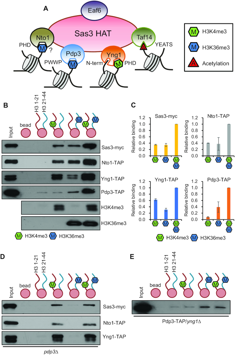
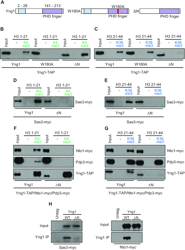
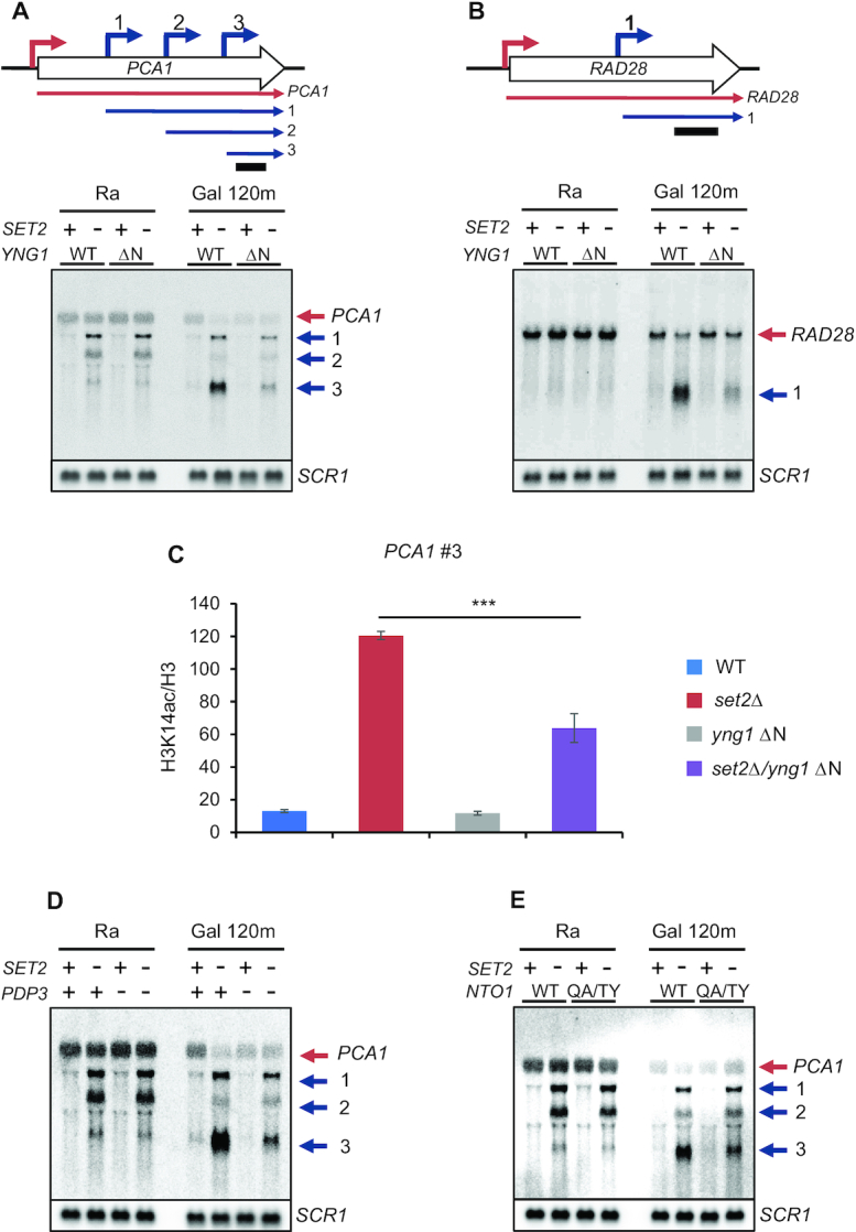
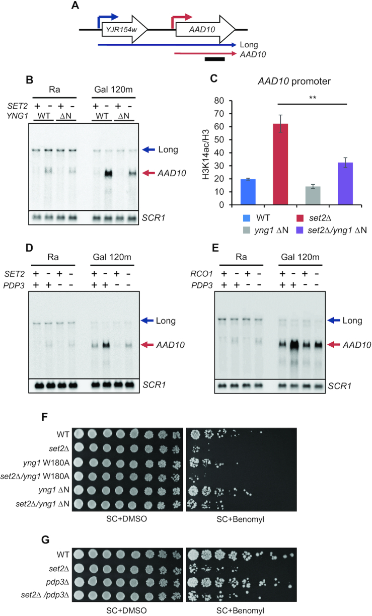
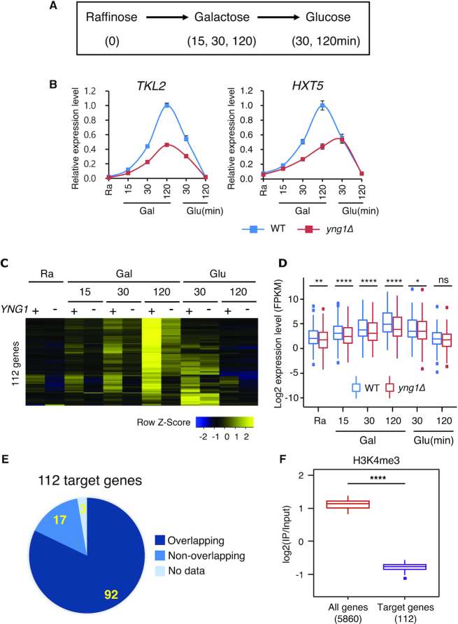
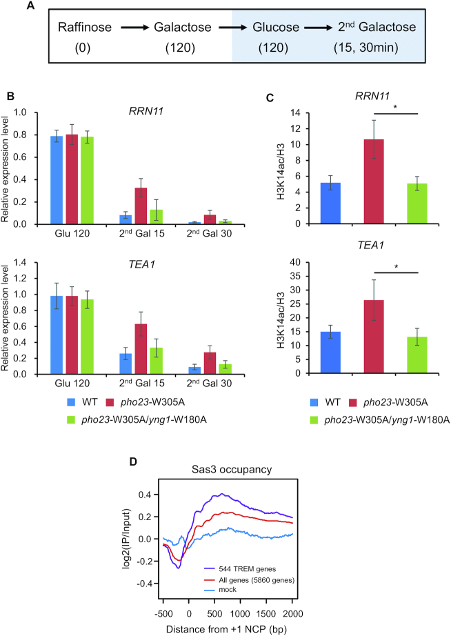
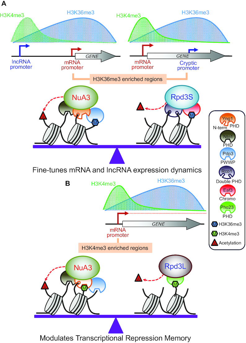
In yeast, NuA3 histone acetyltransferase (NuA3 HAT) promotes acetylation of histone H3 lysine 14 (H3K14) and transcription of a subset of genes through interaction between the Yng1 plant homeodomain (PHD) finger and H3K4me3. Although NuA3 HAT has multiple chromatin binding modules with distinct specificities, their interdependence and combinatorial actions in chromatin binding and transcription remain unknown. Modified peptide pulldown assays reveal that the Yng1 N-terminal region is important for the integrity of NuA3 HAT by mediating the interaction between core subunits and two methyl-binding proteins, Yng1 and Pdp3. We further uncover that NuA3 HAT contributes to the regulation of mRNA and lncRNA expression dynamics by antagonizing the histone deacetylases (HDACs) Rpd3S and Rpd3L. The Yng1 N-terminal region, the Nto1 PHD finger and Pdp3 are important for optimal induction of mRNA and lncRNA transcription repressed by the Set2-Rpd3S HDAC pathway, whereas the Yng1 PHD finger-H3K4me3 interaction affects transcriptional repression memory regulated by Rpd3L HDAC. These findings suggest that NuA3 HAT uses distinct chromatin readers to compete with two Rpd3-containing HDACs to optimize mRNA and lncRNA expression dynamics.


Figures

Figure 1.
Figure 1.
NuA3 HAT exists in several distinct complexes. (A) Schematic representation of multiple chromatin binding domains in NuA3 HAT. Whereas Yng1 PHD finger binds to H3K4me3, its N-terminal region interacts with unmodified histone tails. NuA3 HAT also binds to H3K36me3 via the Pdp3 PWWP domain and the Nto1 PHD finger domain. In addition, the Taf14 YEATS domain is known to bind to histone acetylation. (B) Interdependence and distinct roles of chromatin binding modules of NuA3 HAT. Histone peptide pulldown assays were performed with whole cell extracts from the indicated strains and 1 μg of histone peptides immobilized on magnetic beads in binding buffer containing 250 mM NaCl. Equal amount (0.5 μg of each) of two histone peptides, H3 1–21 and H3 21–44, methylated on K4 or K36 or not was used. Precipitated proteins were analyzed by immunoblot analyses with anti-myc or anti-TAP antibodies. Histone methylation on K4 or K36 was confirmed by immunoblot analyses with anti-H3K4me3 or anti-H3K36me3 antibodies. Two independent experiments showed the same results. (C) Quantitation from (B). Error bars show the standard deviation (S.D.) calculated from two independent experiments. (D) Pdp3 is important for NuA3 HAT binding to H3K36me3. Peptide pulldown assay was done as in (B). Two independent experiments showed the same results. (E) Loss of Yng1 causes dissociation of Pdp3 from H3K4me3. Peptide pulldown assay was performed as in (B). Two independent experiments showed the same results.
Figure 2.
Figure 2.
Yng1 N-terminal region bridges NuA3 HAT core and two methyl-binders. (A) Schematic representation of two chromatin binding domains in Yng1: the Yng1 N-terminal region (amino acids 2–28) and the Yng1 PHD finger (amino acids 141–213). The W180A mutation in PHD finger or deletion of N-terminal region (ΔN) was created by the delitto perfetto strategy. (B) Yng1 binding to H3K4me3 requires its PHD finger but not N-terminal region. Histone peptide pulldown assays were performed with binding buffer containing 300 mM NaCl as in Figure 1B. Two independent experiments showed the same results. (C) The Yng1 N-terminal region is important for association between Yng1 and H3K36me3. Histone peptide pulldown assay was done in binding buffer containing 250 mM NaCl. Two independent experiments showed the same results. (D andE) Interaction between Sas3 and H3K4me3 or H3K36me3 is absent in ΔN strains. Peptide pulldown assay was done as in (C). Two independent experiments showed the same results. (F) Nto1 fails to bind to H3K4me3 and H3K36me3 in the absence of the Yng1 N-terminal region. Peptide pulldown assay was done as in (C). Two independent experiments showed the same results. (G) Only Pdp3 but not Nto1 binds to H3K36me3 in ΔN strains. Peptide pulldown assay was done as in (C). Two independent experiments showed the same results. (H) Loss of the Yng1 N-terminal region disrupts interaction between Yng1 and Sas3 or Nto1. Co-immunoprecipitation assays were carried out whole cell extracts from the indicated strains with Yng1-TAP and IgG beads in binding buffer containing 150 mM NaCl. Precipitated proteins were analyzed by immunoblot analyses with anti-myc or anti-TAP antibodies. Two independent experiments showed the same results.
Figure 3.
Figure 3.
NuA3 HAT positively regulates inducible cryptic promoters repressed by Set2. (A andB) Galactose-inducible cryptic promoters of PCA1 and RAD28 are positively regulated by NuA3 HAT. Northern blot analysis was performed with 3′-strand specific DNA probe. The indicated cells were grown in synthetic complete (SC) medium containing raffinose (Ra) and shifted to SC-galactose media for 120 min (Gal 120m). Bottom panels show cryptic transcripts of PCA1 (A) or RAD28 (B) detected by northern blot analysis, which are schematicized at top. Red arrows are core promoters and blue arrows are cryptic promoters that produce short cryptic transcripts. A bar underneath upper panels indicates position of DNA probe used for northern blot analysis. SCR1 was used as a loading control. Two independent experiments showed the same results. (C) NuA3 HAT acetylates histone H3 at PCA1 cryptic promoter. Cross-linked chromatin from the indicated strains grown in YPD was precipitated with anti-H3 or anti-acetyl H3K14. PCR analysis was carried out on the galactose-inducible cryptic promoter of PCA1. A non-transcribed region near the telomere of chromosome VI was used for an internal control. The signals for acetyl H3K14 were quantitated and normalized to total H3 signal, and the ratios were graphed. Error bars show the standard deviation (S.D.) calculated from three biological replicates, each with three technical replicates. ***P< 0.001 (two-tailed unpaired Student's t tests). (D andE) Pdp3 and Nto1 are required for full activation of galactose-inducible cryptic promoters of PCA1 and RAD28. Northern blot analysis with the indicated strains was done as in (A). Two independent experiments showed the same results.
Figure 4.
Figure 4.
NuA3 HAT antagonizes the Set2-Rpd3S pathway. (A) Blue and red arrow indicate a distal promoter that produces a lncRNA and a core promoter for AAD10 mRNA, respectively. A bar underneath indicates position of probe used for northern blot analysis. (B) Loss of the Yng1 N-terminal region attenuates AAD10 induction in SET2 deleting cells. Northern blot analysis of AAD10 was performed with a 3′-strand specific DNA probe. The indicated cells were grown in synthetic complete (SC) medium containing raffinose (Ra) and shifted to SC-galactose media for 120 min (Gal 120m). SCR1 was used as a loading control. Two independent experiments showed the same results. (C) NuA3 HAT acetylates histones at the AAD10 promoter. ChIP assay was performed as in Figure 3C. **P< 0.01 (two-tailed unpaired Student's t tests). (D andE) Pdp3 is required for AAD10 induction in mutants for the Set2-Rpd3S pathway. Northern blot analysis of AAD10 was carried out as in (B). (F) Loss of the Yng1 N-terminal region but not its PHD finger mutation partially suppresses the growth defect of SET2 deleting cells in the presence of Benomyl. The indicated strains were spotted in 3-fold dilutions on synthetic complete (SC) medium containing DMSO (2 days growth shown) or 50 μg/ml Benomyl (5.6 days growth shown). (G) Growth defect of set2Δ is partially suppressed by deletion of PDP3. Spot assay of the indicated strains was done as in (F).
Figure 5.
Figure 5.
NuA3 HAT fine-tunes the kinetics of transcriptional induction. (A) Schematic representation of the time course experiments to monitor changes in transcript levels upon carbon source shifts. (B) RNA samples from the time course experiments in (A) were analyzed by RT-PCR. SCR1 was used as an internal control. Error bars show the standard deviation (S.D.) calculated from two biological replicates, each with three technical replicates. (C) Two RNA samples from the time course experiments in (B) were analyzed by strand-specific RNA sequencing. NuA3 HAT-activated genes were identified as those showing at least 1.7-fold decrease in transcript levels at one more time points and the P-value from Cuffdiff < 0.05. (D) Averaged profiles of expression signals of 112 genes in (C); ns: not significant, *P < 0.05, **P < 0.01, ***P < 0.001, and ****P < 0.0001 (paired t-test). (E) A pie chart shows the number of genes (yellow) with overlapping lncRNA transcription. Among these, 82% (92 genes) of genes are overlapped with lncRNA transcription. (F) H3K4me3 is depleted at promoters of NuA3 HAT target genes. The average enrichment of H3K4me3 for all genes (red) and for 112 genes from (C; purple). H3K4me3 pattern was analyzed using the data sets from Weiner et al. (34). ****P < 0.0001 (paired t-test).
Figure 6.
Figure 6.
Interaction between the Yng1 PHD finger and H3K4me3 is important for TREM. (A) Schematic representation of the time course experiments to monitor changes in transcript levels upon carbon source shifts. (B) Loss of the Yng1 PHD finger–H3K4me3 interaction facilitates gene repression in mutant for PHO23. RNA samples at glucose 120 min (Glu 120), second galactose 15 min (second Gal 15) and second galactose 30 min (second Gal 30) from (A) were analyzed by RT-PCR. SCR1 was used as an internal control. Error bars show the standard deviation (S.D.) calculated from two biological replicates, each with three technical replicates. (C) NuA3 HAT acetylates histones at the promoters of TREM genes. ChIP assay was performed as in Figure 3C. Error bars show the standard deviation (S.D.) calculated from three biological replicates, each with three technical replicates. *P < 0.05 (two-tailed unpaired Student’s t tests). (D) NuA3 binds to TREM genes. The average enrichment of Sas3 occupancy relative to the +1 nucleosome core particle (NCP) position analyzed using the data sets from Martin et al. (22). The plots represent the average enrichment of Sas3-HA for all genes (red) and for 544 TREM genes (purple). Blue indicates the average of log2 (IP/Input) values from mock sample.
Figure 7.
Figure 7.
Models for regulation of gene expression by NuA3 HAT complex. (A) Opposing roles of NuA3 HAT and Rpd3S HDAC in regulation of histone acetylation at H3K36me3-enriched promoters. Transcription from an upstream lncRNA promoter targets H3K36me3 at the core promoter of mRNA target genes. In addition, transcription from the mRNA promoter places H3K36me3 in 3′ transcribed regions. Pdp3 of NuA3 HAT or Eaf3 chromodomain of Rpd3S HDAC modulates histone acetylation. The Yng1 N-terminal region, Nto1, and Rco1 may interact with unmodified histone tails to stabilize chromatin binding of NuA3 HAT or Rpd3S HDAC. Antagonistic function of NuA3 HAT and Rpd3S HDAC fine-tunes mRNA and lncRNA expression dynamics upon environmental changes. (B) NuA3 HAT may compete with Rpd3L HDAC at H3K4me3-enriched promoters to regulate histone acetylation and TREM. The Yng1 PHD finger of NuA3 HAT or Pho23 PHD finger of Rpd3L HDAC binds to H3K4me3 to control histone acetylation. Optimal level of histone acetylation at H3K4me3-enriched promoters is important for regulation of TREM.

Similar articles

Cited by

References

    1. Kouzarides T. Chromatin modifications and their function. Cell. 2007; 128:693–705. - PubMed
    1. Li B., Carey M., Workman J.L.. The role of chromatin during transcription. Cell. 2007; 128:707–719. - PubMed
    1. Buratowski S., Kim T.. The role of cotranscriptional histone methylations. Cold Spring Harb. Symp. Quant. Biol. 2010; 75:95–102. - PMC - PubMed
    1. Woo H., Dam Ha S., Lee S.B., Buratowski S., Kim T.. Modulation of gene expression dynamics by co-transcriptional histone methylations. Exp. Mol. Med. 2017; 49:e326. - PMC - PubMed
    1. Shi X., Kachirskaia I., Walter K.L., Kuo J.H., Lake A., Davrazou F., Chan S.M., Martin D.G., Fingerman I.M., Briggs S.D. et al. .. Proteome-wide analysis in Saccharomyces cerevisiae identifies several PHD fingers as novel direct and selective binding modules of histone H3 methylated at either lysine 4 or lysine 36. J. Biol. Chem. 2007; 282:2450–2455. - PMC - PubMed

Publication types

MeSH terms