Review
. 2020 Nov 18;48(20):11227-11243.
doi: 10.1093/nar/gkaa777.

The involvement of nucleotide excision repair proteins in the removal of oxidative DNA damage

Affiliations
Review

The involvement of nucleotide excision repair proteins in the removal of oxidative DNA damage

Namrata Kumar et al. Nucleic Acids Res. .

Abstract

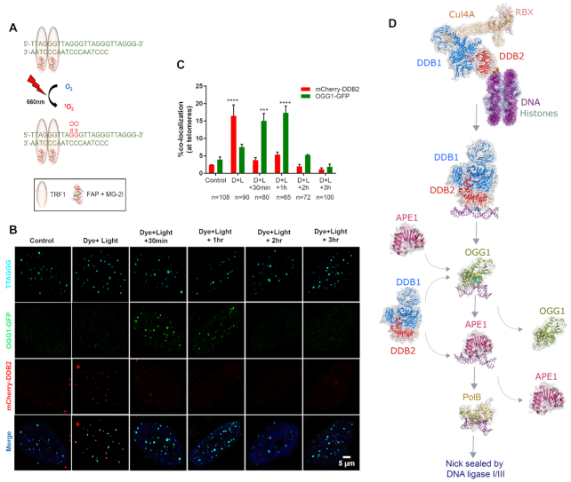
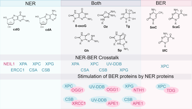
The six major mammalian DNA repair pathways were discovered as independent processes, each dedicated to remove specific types of lesions, but the past two decades have brought into focus the significant interplay between these pathways. In particular, several studies have demonstrated that certain proteins of the nucleotide excision repair (NER) and base excision repair (BER) pathways work in a cooperative manner in the removal of oxidative lesions. This review focuses on recent data showing how the NER proteins, XPA, XPC, XPG, CSA, CSB and UV-DDB, work to stimulate known glycosylases involved in the removal of certain forms of base damage resulting from oxidative processes, and also discusses how some oxidative lesions are probably directly repaired through NER. Finally, since many glycosylases are inhibited from working on damage in the context of chromatin, we detail how we believe UV-DDB may be the first responder in altering the structure of damage containing-nucleosomes, allowing access to BER enzymes.


Figures

Figure 1.
Figure 1.
Chemical structures of oxidative lesions formed in DNA. (A) Various oxidation products of guanine. (B) Formation of cyclic guanosine by oxidation. (C) Formation of cyclic adenosine by oxidation. (D) Enzymatic oxidative demethylation of 5-methylcytosine. (E) Oxidation of thymine to thymine glycol. ROS produces over 100 different types of lesions in DNA, and this figure displays the structures of those damages that are discussed in this review. ROS: Reactive oxygen species; DNMT: DNA methyltransferase; TET: Ten-eleven translocation enzymes.
Figure 2.
Figure 2.
Mammalian base excision repair (BER) pathway. The base lesion is excised by a lesion-specific DNA glycosylase. Monofunctional glycosylases break the glycosidic bond between the damaged base and the sugar moiety, resulting in an abasic site. AP endonuclease 1 (APE1) processes the abasic site to form a 3′OH and a deoxyribose-5′-phosphate (dRP), which is removed by the lyase activity of DNA polymerase β (pol β). Bifunctional glycosylases utilize their AP lyase activity to cleave the phosphate backbone, creating a single strand break, leaving a free 5′ phosphate and either a 3′-phospho-α, β-unsaturated aldehyde (3′-PUA) (β-elimination) or a 3′ phosphate (β,δ-elimination). APE1 acts on the β-elimination product while polynucleotide kinase phosphate (PNKP) is required to process the 3′phosphate after β,δ-elimination. The resulting 3′OH is bound by PARP1 which recruits the BER complex consisting of pol β, XRCC1 and DNA ligase. The one base gap is then filled by pol β and the nick in the DNA is sealed by DNA ligase. Adapted from Kumar et al. (8) with permission.
Figure 3.
Figure 3.
BER glycosylases, their structures and respective substrates. The glycosylases (green) are bound to DNA (purple) containing a lesion (purple, space-filled). All structures are human except SMUG1 (Xenopus laevis), MUTYH (Geobacillus stearothermophilus), NEIL2 (Monodelphis domestica), NEIL3 (Mus musculus), NTHL1 (EndoIII, Geobacillus stearothermophilus). PDB: SMUG1 (1OE4), MBD4 (5CHZ), UNG (1EMH), TDG (3UFJ), MPG (1BNK), MUTYH (4YOQ), OGG1 (1EBM), NEIL1 (5ITY), NEIL2 (6VJI), NEIL3 (3W0F), NTHL1 (1ORN). Abbreviations: U, uracil; A, adenine; T, thymine; C, cytosine; G, guanine; 5-FU, 5-fluorouracil; 5-hmU, 5-hydroxymethyluracil; ϵ, etheno; FaPy, 2,6-diamino-4-hydroxy-5-N-methylformamidopyrimidine; 8-oxoG, 8-oxoguanine; Gh, Guanidonohydantoin; Sp, Spiroiminodihydantonin; Im, iminoallantoin; 5fC, 5-formylcytosine; 5caC, 5-carboxycytosine; 5-BrU, 5-Bromouracil; Tg, Thymine Glycol; meA, 3-methyladenine; meG, 3-methylguanine; 5-hC, 5-hydroxycytosine; 5-hU, 5-hydroxyuracil; 2-hA, 2-hydroxyadenine
Figure 4.
Figure 4.
XPC and TDG in oxidative demethylation of 5-methylcytosine. Based on the work by Ho et al. (60), XPC works to help turnover TDG, which like other glycosylases, is product inhibited binding tightly to abasic sites. Shown here is the structure of the yeast, XPC homolog, Rad4 (green) Rad23 (red) bound to a DNA duplex (purple) containing a 6–4 photoproduct (blue space-filled), PDB: 6CFI; Human TDG (green) bound to a DNA duplex (purple) containing 2′-fluoro-2′-deoxyuridine (blue space-filled), PDB:3UFJ (122).
Figure 5.
Figure 5.
Role of UV-DDB in 8-oxoguanine (8-oxoG) repair. (A) A chemoptogenetic approach to introduce 8-oxoG at telomeres. Fluorogen-activating peptide (FAP) is fused to a telomere binding protein (TRF1). In the presence of a malachite green dye (MG2I), the FAP-TRF1-MG2I combination is excitable at far red wavelength (660nm) and generates singlet oxygen. Singlet oxygen reacts with telomeric DNA to form 8-oxoG lesions. (B) Immunofluorescence images of mCherry-DDB2 (red) and OGG1-GFP (green) recruitment to and departure from 8-oxoG lesions at telomeres (blue). (C) Quantitative analysis of immunofluorescence images in (B). (D) Working model of the potential role of UV-DDB in BER of 8-oxoG. Figure adapted from Jang et al. (56) with permission. Model of human UV-DDB-CUL4A-RBX bound to a 6–4 photoproduct in the context of a nucleosome, built from PDB codes: 4A0K and 6R8Y (52,123). Human UV-DDB bound to a THF lesion, PDB: 4E54 (124). Human OGG1 bound to 8-oxoG, PDB: 1EBM (125). Human APE1 bound to a 3′ deoxyribose phosphate moiety, PDB: 5DFF (126). DNA polymerase beta bound to gapped and nicked DNA, PDB: 1BPX (127)
Figure 6.
Figure 6.
Oxidative DNA damage lesions repaired by BER, NER or both. NER proteins involved in the removal of these oxidative lesions are highlighted. Also depicted above is stimulation of BER proteins, especially glycosylases, by NER proteins. In this schematic, NER refers to both global and transcription-coupled repair. cdG: cyclo-deoxyguanosine; cdA: cyclo-deoxyadenosine; 8-oxoG: 8-oxoguanine; Oz: Oxazolone; Gh: Guanidinohydantoin; Sp: Spiroiminodihydantoin; Tg: Thymine glycol; 5mC: 5-methylcytosine; 5fC: 5-formylcytosine; 5caC: 5-carboxycytosine.

References

    1. Markkanen E. Not breathing is not an option: how to deal with oxidative DNA damage. DNA Repair (Amst.). 2017; 59:82–105. - PubMed
    1. Reuter S., Gupta S.C., Chaturvedi M.M., Aggarwal B.B.. Oxidative stress, inflammation, and cancer: how are they linked. Free Radic. Biol. Med. 2010; 49:1603–1616. - PMC - PubMed
    1. Tubbs A., Nussenzweig A.. Endogenous DNA damage as a source of genomic instability in cancer. Cell. 2017; 168:644–656. - PMC - PubMed
    1. Poetsch A.R. The genomics of oxidative DNA damage, repair, and resulting mutagenesis. Comput. Struct. Biotechnol. J. 2020; 18:207–219. - PMC - PubMed
    1. Sedelnikova O.A., Redon C.E., Dickey J.S., Nakamura A.J., Georgakilas A.G., Bonnera W.M.. Role of oxidatively induced DNA lesions in human pathogenesis. Mutat. Res. 2010; 704:152–159. - PMC - PubMed

Publication types