Review
. 2021 Jan;191(1):26-39.
doi: 10.1016/j.ajpath.2020.09.006. Epub 2020 Oct 1.

The Origin of Ovarian Cancer Species and Precancerous Landscape

Affiliations
Review

The Origin of Ovarian Cancer Species and Precancerous Landscape

Ie-Ming Shih et al. Am J Pathol. 2021 Jan.

Abstract

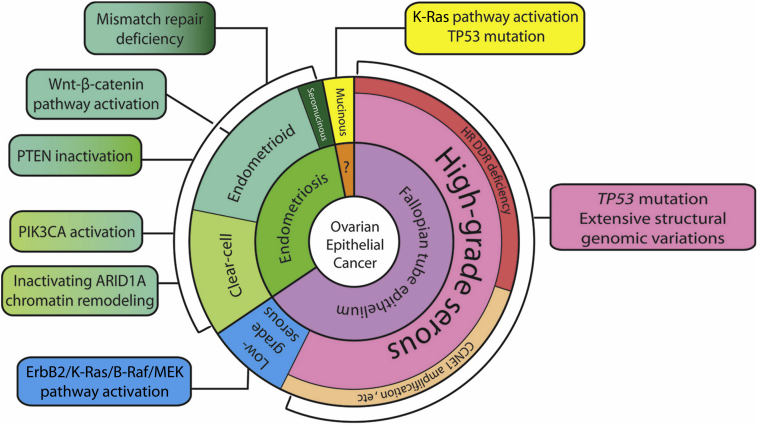
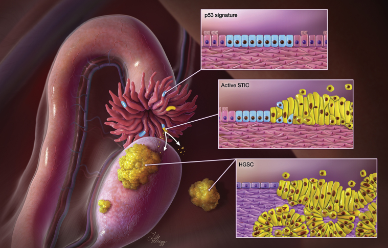
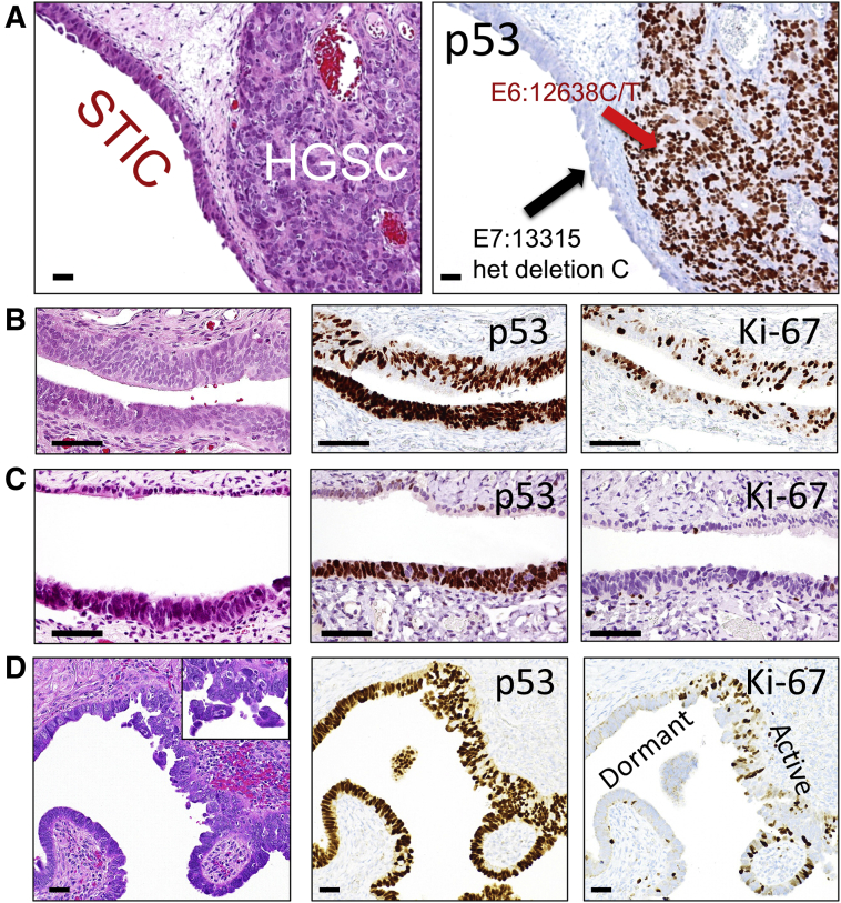
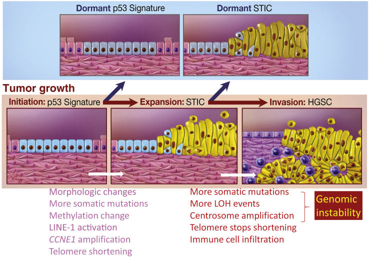
Unlike other human cancers, in which all primary tumors arise de novo, ovarian epithelial cancers are primarily imported from either endometrial or fallopian tube epithelium. The prevailing paradigm in the genesis of high-grade serous carcinoma (HGSC), the most common ovarian cancer, posits to its development in fallopian tubes through stepwise tumor progression. Recent progress has been made not only in gathering terabytes of omics data but also in detailing the histologic-molecular correlations required for looking into, and making sense of, the tissue origin of HGSC. This emerging paradigm is changing many facets of ovarian cancer research and routine gynecology practice. The precancerous landscape in fallopian tubes contains multiple concurrent precursor lesions, including serous tubal intraepithelial carcinoma (STIC), with genetic heterogeneity providing a platform for HGSC evolution. Mathematical models imply that a prolonged time (decades) elapses from the development of a TP53 mutation, the earliest known molecular alteration, to an STIC, followed by a shorter span (6 years) for progression to an HGSC. Genetic predisposition accelerates the trajectory. This timeline may allow for the early diagnosis of HGSC and STIC, followed by intent-to-cure surgery. This review discusses the recent advances in this tubal paradigm and its biological and clinical implications, alongside the promise and challenge of studying STIC and other precancerous lesions of HGSC.


Figures

Figure 1
Figure 1
The tissue origins and major molecular pathway alterations in different types of ovarian epithelial cancer. ARID, AT-rich interactive domain-containing protein; CCNE, G1/S-specific cyclin-E; ErbB, extracellular region binding protein; HR DDR, homologous recombination DNA damage repair; MEK (alias mitogen-activated protein kinase, MAPK); PIK3CA, phosphatidylinositol 3-kinase catalytic subunit α; PTEN, phosphatase and tensin homologue.
Figure 2
Figure 2
Paradigm of fallopian tube as the origin of high-grade serous carcinomas (HGSCs). Multiple fallopian tube lesions including serous tubal intraepithelial carcinoma (STIC) (yellow lines in the fimbriated end) and p53 signature (blue lines) can occur at the fimbriated end. STIC is presumed to be the immediate precursor of HGSC. STIC cells can become invasive in the fallopian tube, and detach from the fallopian tube surface, spreading onto the peritoneal surface, enclosing ovary, bowel, peritoneal wall, and omentum. Natural selection favors emigrated STIC cells that can survive and reproduce within a certain tissue-environmental niche which grow into tumor nodules and cause tumor ascites. Illustration by Lydia Gredd, M.A., C.M.I., © 2020 I. Shih at JHU; used with permission.
Figure 3
Figure 3
Examples of serous tubal intraepithelial carcinoma (STIC) and high-grade serous carcinoma (HGSC). A: A STIC is closely associated with an invasive HGSC. Both lesions contain different TP53 mutations, suggesting that the HGSC is clonally independent of the STIC. B: An example of STIC with an intense and diffuse p53 staining pattern compatible with a missense TP53 mutation and a high proliferative activity as indicated by Ki-67–labeled epithelial cells. C: A dormant STIC with a TP53 mutation staining pattern of a very low proliferative activity. D: Another example of a STIC showing both proliferation active and dormant STIC areas. Hematoxylin and eosin stain sections of B, C, and D are shown in the left panels. The inset in D shows loose clusters of STIC cells detaching from the tubal surface. Scale bars = 100 μm (AD). Original magnification, ×20 (inset).
Figure 4
Figure 4
Molecular alterations associated with tumor progression from p53 signature to serous tubal intraepithelial carcinoma (STIC) to high-grade serous carcinoma (HGSC). After initial clonal expansion, p53 signatures and some STICs become dormant as they lose a proliferative advantage. But some STICs continue to proliferate, gain additional cancer-promoting events, and progress to HGSC spreading to fallopian tubes, ovaries, and other pelvic organs. Compared to p53 signatures, STICs gain several features as listed, and may be involved in the progression. Similarly, as compared to STICs, HGSCs are more frequently characterized by features related to genomic instability, maintenance of telomere length, and immune cell infiltration. LINE, long-interspersed element; LOH, loss of heterozygosity.
Figure 5
Figure 5
Alternative pathways in the development of high-grade serous carcinoma (HGSC). A: HGSC arising from an ovarian serous cystadenoma. Top left panel: A low-magnification view showing the wall of the cystadenoma with a focal solid growth of HGSC. Top right panel: Normal-appearing epithelium surrounding the solid tumor. Bottom panel: Histologic features of an HGSC. B: HGSC developing directly on the ovarian surface. Top left panel: A low-magnification view showing the whole ovarian section. Top right panel: The ovarian surface showing adenofibroma where the HGSC arises. Bottom panel: HGSC on the surface of adenofibroma. C: HGSC arising from an enlarged ovarian cortical inclusion cyst. Top panel: A low-magnification view of the inclusion cyst. Bottom panel: Highly atypical cells appearing on the surface of the cyst. Scale bars: 100 μm (A, top right panel and bottom panel; B, bottom panel; C, bottom panel); 1 mm (B, top right panel; C, top panel); 1 cm (A, top left panel; B, top left panel).

Similar articles

Cited by

References

    1. Kurman R.J., Shih I.M. The origin and pathogenesis of epithelial ovarian cancer: a proposed unifying theory. Am J Surg Pathol. 2010;34:433–443. - PMC - PubMed
    1. Kurman R.J., Shih IeM. The Dualistic model of ovarian carcinogenesis: revisited, revised, and expanded. Am J Pathol. 2016;186:733–747. - PMC - PubMed
    1. Shih IeM., Panuganti P.K., Kuo K.T., Mao T.L., Kuhn E., Jones S., Velculescu V.E., Kurman R.J., Wang T.L. Somatic mutations of PPP2R1A in ovarian and uterine carcinomas. Am J Pathol. 2011;178:1442–1447. - PMC - PubMed
    1. Maeda D., Shih IeM. Pathogenesis and the role of ARID1A mutation in endometriosis-related ovarian neoplasms. Adv Anat Pathol. 2013;20:45–52. - PMC - PubMed
    1. Kuhn T.S., Hacking I. ed 4. The University of Chicago Press; Chicago: 2012. The Structure of Scientific Revolutions.

Publication types

MeSH terms