. 2020 Dec:42:101091.
doi: 10.1016/j.molmet.2020.101091. Epub 2020 Oct 1.

β-catenin regulates muscle glucose transport via actin remodelling and M-cadherin binding

Affiliations

β-catenin regulates muscle glucose transport via actin remodelling and M-cadherin binding

Stewart W C Masson et al. Mol Metab. 2020 Dec.

Abstract

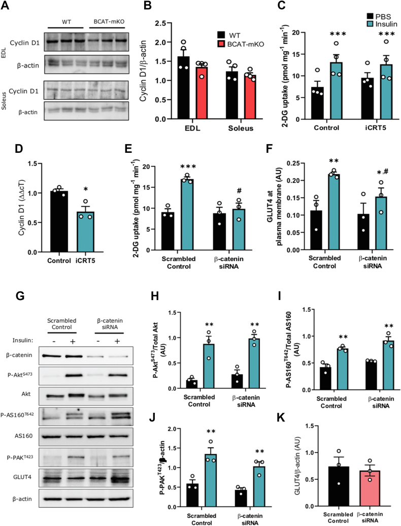
Objective: Skeletal muscle glucose disposal following a meal is mediated through insulin-stimulated movement of the GLUT4-containing vesicles to the cell surface. The highly conserved scaffold-protein β-catenin is an emerging regulator of vesicle trafficking in other tissues. Here, we investigated the involvement of β-catenin in skeletal muscle insulin-stimulated glucose transport.

Methods: Glucose homeostasis and transport was investigated in inducible muscle specific β-catenin knockout (BCAT-mKO) mice. The effect of β-catenin deletion and mutation of β-catenin serine 552 on signal transduction, glucose uptake and protein-protein interactions were determined in L6-G4-myc cells, and β-catenin insulin-responsive binding partners were identified via immunoprecipitation coupled to label-free proteomics.

Results: Skeletal muscle specific deletion of β-catenin impaired whole-body insulin sensitivity and insulin-stimulated glucose uptake into muscle independent of canonical Wnt signalling. In response to insulin, β-catenin was phosphorylated at serine 552 in an Akt-dependent manner, and in L6-G4-myc cells, mutation of β-cateninS552 impaired insulin-induced actin-polymerisation, resulting in attenuated insulin-induced glucose transport and GLUT4 translocation. β-catenin was found to interact with M-cadherin in an insulin-dependent β-cateninS552-phosphorylation dependent manner, and loss of M-cadherin in L6-G4-myc cells attenuated insulin-induced actin-polymerisation and glucose transport.

Conclusions: Our data suggest that β-catenin is a novel mediator of glucose transport in skeletal muscle and may contribute to insulin-induced actin-cytoskeleton remodelling to support GLUT4 translocation.

Keywords: Actin-remodelling; Beta-catenin; GLUT4 trafficking; Glucose transport; M-cadherin.


Figures

Image 1
Graphical abstract
Figure 1
Figure 1
Depletion of β-catenin in skeletal muscle of adult mice mildly impairs glucose homeostasis without affecting tissue masses or energy expenditure. One month following tamoxifen (TX) treatment, β-catenin expression in gastrocnemius (gastroc), extensor digitorum longus (EDL), and soleus muscle (A–B), body mass (C), body composition (D), food and water intake (E–F), energy expenditure (G), respiratory exchange ratio (H), glucose tolerance (I–J), as well as fed and fasted blood glucose (K) and plasma insulin (L) were determined in WT and BCAT-mKO mice. Results are mean ± SE, with individual animals shown as data points in figures or in figure legend. Significance was determined using two-tailed Student's t-test (J), two-way ANOVA with LSD post-hoc analysis (B, D–H, K–L) or two-way RM ANOVA with LSD post-hoc analysis (C, I). TA, tibialis anterior muscle; sub, subcutaneous fat pad; epi, epididymal fat pad; BAT, brown adipose tissue. ∗∗∗p < 0.001 significance within groups.
Figure 2
Figure 2
Reduced β-catenin in skeletal muscle of adult mice causes insulin resistance and impaired insulin-induced skeletal muscle glucose transport. One month following tamoxifen (TX) treatment, insulin tolerance was measured (A–B) before assessment of in vivo insulin-stimulated glucose transport into the gastrocnemius, tibialis anterior, subcutaneous and epidydimal fat pads of BCAT-mKO and WT mice (C–F). Isolated muscle insulin-stimulated glucose transport was determined in EDL (extensor digitorium longus) and soleus muscle from WT and BCAT-mKO mice (G–H). Akt phosphorylation and GLUT4 protein expression in the gastrocnemius muscles of BCAT-mKO and WT mice was assessed by immunoblot (I–K). Results are mean ± SE, with individual animals shown as data points in figures or in figure legend. Significance was determined using two-tailed Student's t-test (B, K), two-way ANOVA with LSD post-hoc analysis (C–F, J) or two-way RM ANOVA with LSD post-hoc analysis (G–H). ∗p < 0.05, ∗∗p < 0.01, ∗∗∗p < 0.001 significance within groups. #p < 0.05 significance between groups. AUC, area under curve.
Figure 3
Figure 3
Depletion of β-catenin in L6-G4-myc cells impairs insulin-stimulated glucose transport and actin remodelling independent of Wnt-mediated transcription. Cyclin D1 protein expression in gastrocnemius muscle of BCAT-mKO and WT muscle was assessed by immunoblot (A–B). Glucose uptake and analysis of cyclin D1 mRNA expression in L6-G4-myc myoblasts treated with iCRT5 (50 μM) for 1 h (C–D). L6-G4-myc myoblasts were treated with β-catenin specific siRNA prior to assessment of glucose uptake (E–F) and insulin-signalling pathway (G–K). Results are mean ± SE, with individual animals or biological replicates shown as data points in figures or in figure legend. Significance was determined using two-tailed Student's t-test (D, K) or two-way ANOVA with LSD post-hoc analysis (B, C, E–J). ∗p < 0.05, ∗∗∗p < 0.001 significance within groups. #p < 0.05 significance between groups.
Figure 4
Figure 4
Insulin phosphorylates β-cateninS552downstream of proximal insulin signalling, and β-cateninS552phosphorylation is required for insulin-stimulated glucose transport. Immunoblotting analysis of P-β-cateninS552, β-catenin, P-AktS473 Akt, and β-actin in C57Bl/6j mice treated with either glucose or insulin prior to tissue collection (A–B). Immunoblotting analysis of P-β-cateninS552, β-catenin, P-AktS473, Akt and β-actin in L6-G4-myc cells incubated with either BYL719 or AKTi-1/2 prior to insulin stimulation and (C–E). Insulin-stimulated glucose uptake and GLUT4 translocation to the membrane in L6-G4-myc cells transfected with S552A-β-catenin (F–H). Results are mean ± SE, with individual animals or biological replicates shown as individual data points in figures or in figure legend. Significance was determined using, one (B) or two-way ANOVA with LSD post-hoc analysis (D, E and G, H). ∗p < 0.05, ∗∗p < 0.01 significance within groups. #p < 0.05 significance between groups.
Figure 5
Figure 5
Insulin promotes β-catenin/M-cadherin interaction via β-cateninS552phosphorylation. L6-G4-myc myotubes were treated with either insulin or PBS (control) prior to lysis and immunoprecipitation with β-catenin-specific antibody (A). L6-G4-myc myoblasts were treated with M-cadherin-specific siRNA prior to determination of F-actin % (B) and glucose uptake (C) following insulin stimulation. F actin % in response to insulin (D) and PDGF-mediated glucose uptake (E) and immunoblotting of M-cadherin following immunoprecipitation with anti-β-catenin antibody (F–G) in S552A-β-catenin expressing L6-G4-myc myoblasts. Results are mean ± SE, with biological replicates shown as individual data points in figures or in figure legend. Significance was determined using the sign-rank test (A) or two-way ANOVA with LSD post-hoc analysis (B–G). ∗p < 0.05, ∗∗p < 0.01, ∗∗∗p < 0.001 significance within groups. #p < 0.05 significance between groups.
Figure S1
Figure S1
Figure S2
Figure S2
Figure S3
Figure S3

Similar articles

Cited by

References

    1. DeFronzo R.A., Ferrannini E. Regulation of hepatic glucose metabolism in humans. Diabetes/Metabolism Research and Reviews. 1987;3(2):415–459. - PubMed
    1. Mîinea C.P., Sano H., Kane S., Sano E., Fukuda M., Peränen J. AS160, the Akt substrate regulating GLUT4 translocation, has a functional Rab GTPase-activating protein domain. Biochemical Journal. 2005;391(1):87–93. - PMC - PubMed
    1. Wang Q., Somwar R., Bilan P.J., Liu Z., Jin J., Woodgett J.R. Protein kinase B/Akt participates in GLUT4 translocation by insulin in L6 myoblasts. Molecular and Cellular Biology. 1999;19(6):4008–4018. - PMC - PubMed
    1. Tunduguru R., Zhang J., Aslamy A., Salunkhe V.A., Brozinick J.T., Elmendorf J.S. The actin-related p41ARC subunit contributes to p21-activated kinase-1 (PAK1)-mediated glucose uptake into skeletal muscle cells. Journal of Biological Chemistry. 2017:801340. M117. - PMC - PubMed
    1. Tunduguru R., Chiu T.T., Ramalingam L., Elmendorf J.S., Klip A., Thurmond D.C. Signaling of the p21-activated kinase (PAK1) coordinates insulin-stimulated actin remodeling and glucose uptake in skeletal muscle cells. Biochemical Pharmacology. 2014;92(2):380–388. - PMC - PubMed

Publication types

MeSH terms