. 2021 Feb 15;89(4):376-385.
doi: 10.1016/j.biopsych.2020.08.008. Epub 2020 Aug 18.

Long-Term Cocaine Self-administration Produces Structural Brain Changes That Correlate With Altered Cognition

Affiliations

Long-Term Cocaine Self-administration Produces Structural Brain Changes That Correlate With Altered Cognition

Hank P Jedema et al. Biol Psychiatry. .

Abstract

Background: An enduring question from cross-sectional clinical studies is whether the structural and functional differences often observed between cocaine users and healthy control subjects result from a history of drug use or instead reflect preexisting differences. To assess causality from drug exposure, true predrug baseline imaging and neurocognitive assessments are needed.

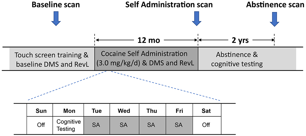
Methods: We addressed this fundamental question of causality using longitudinal anatomical magnetic resonance imaging and neurocognitive assessments in rhesus macaques. Cognitive tasks employed were stimulus reversal learning as a measure of cognitive flexibility/inhibitory control and delayed match to sample as a measure of visual working memory. Time points examined were before and following 12 months of chronic cocaine (n = 8) or water (n = 6) self-administration. A magnetic resonance imaging-only time point was also obtained following 2 years of forced abstinence.

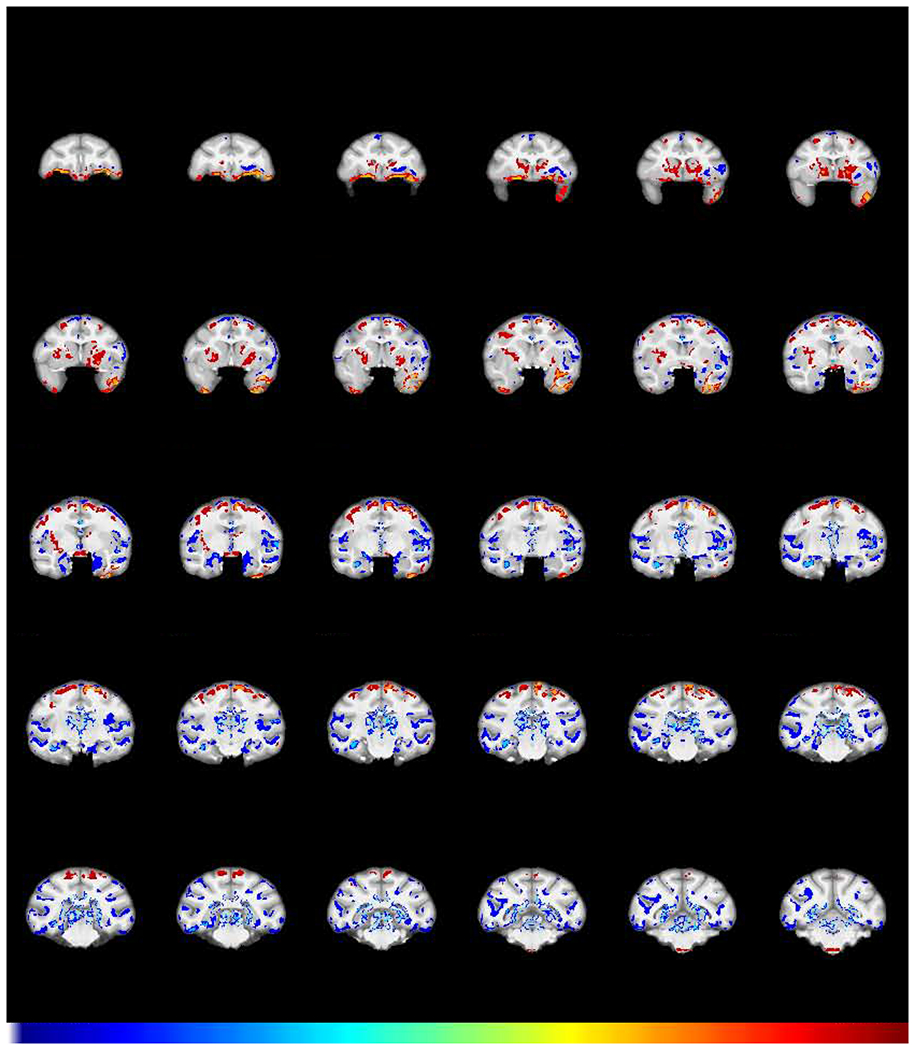
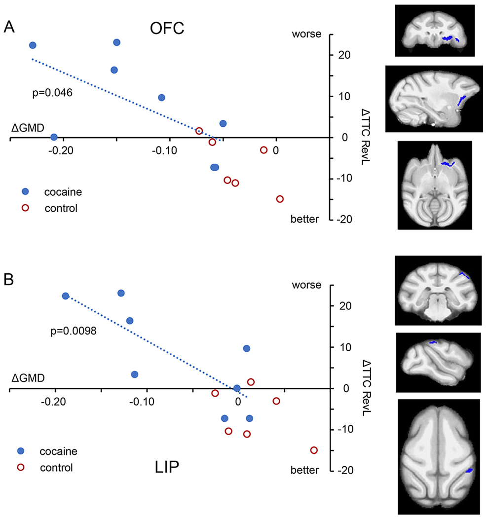
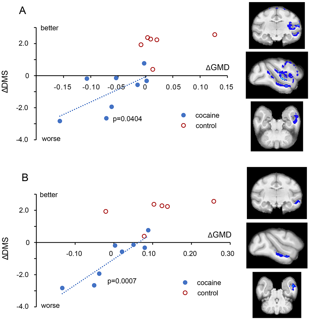
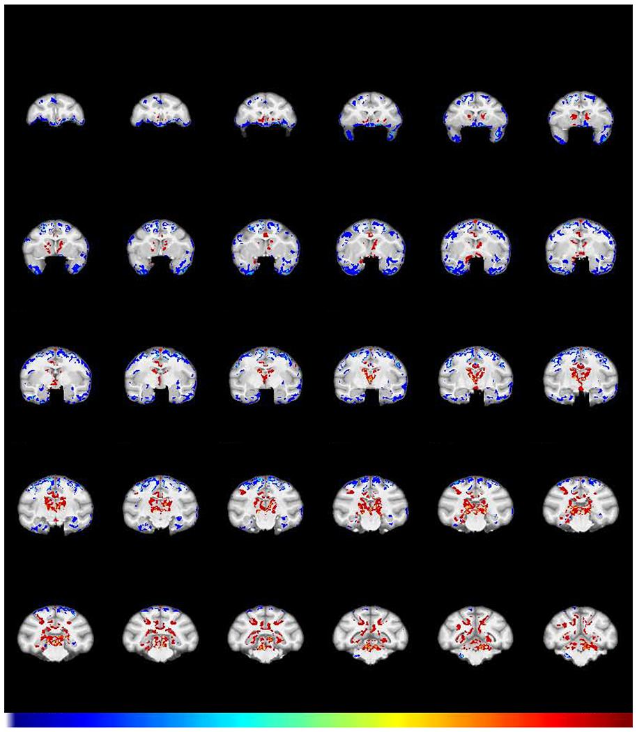
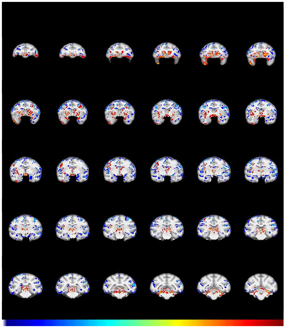
Results: We identified localized patterns of gray matter density (GMD) changes that were largely concordant with cross-sectional clinical studies. These included decreases in orbitofrontal cortex, insula, amygdala, and temporal cortex. There was also a prominent increase in GMD in the caudate putamen. GMD decreases were significantly correlated with cognitive impairments across individuals only in select cortical regions. Following abstinence, changes in GMD in some regions, including the orbitofrontal cortex, insula, and amygdala, were persistent and thus may play an important role in risk of relapse following extended abstinence.

Conclusions: Cocaine use is causal in producing regional changes in GMD, and those changes appear to drive cognitive impairments.

Keywords: Addiction; Cocaine; Cognition; Impairment; Macaque; Structural imaging.


Conflict of interest statement

Financial disclosures: The authors report no biomedical financial interests or potential conflicts of interest.

Figures

Fig 1
Fig 1
Experimental timeline. Subjects received a structural MRI scan and were trained on a touch screen to perform a Delayed Match to Sample (DMS) task and a stimulus discrimination with reversal learning (RevL) task prior to assignment into one of two groups (cocaine self-administration (N=8), water self-administration (SA) control (N=6)). Cocaine animals self-administered intravenously up to 3.0mg/kg cocaine 4 days a week, while control subjects performed similar tasks for water reward. Cognitive performance on DMS and RevL tasks were assessed on alternating Mondays.
Fig 2
Fig 2
Effect of chronic cocaine on GMD. Coronal sections of structural interaction resulting from cocaine self-administration (FWE-corrected −log(p) TFCE maps showing the difference between groups in GMD change from baseline to post-self-administration scans). Regions in which cocaine subjects (N=8) exhibited a greater relative decline in GMD compared to control subjects (N=6) are colored in blue. Regions in which the cocaine subjects showed relatively greater increases in GMD are colored in red. Numbers above sections represent distance in mm from the crossing of the anterior commissure.
Fig 3
Fig 3
Impact of structural changes on RevL performance. A. Within the cocaine subjects (blue symbols), the impairment in RevL performance (greater number of trials to criterion) correlated with the magnitude of GMD decline in regions of the orbitofrontal cortex (OFC; 471 voxels). B. The lateral parietal area (LIP; 187 voxels) also showed a significant correlation. Control subjects (red) are shown for comparison. Orthogonal views (coronal, parasagittal, and axial) indicate the location of the region of decreased GMD. For all regressions, only cocaine subjects were used.
Fig 4
Fig 4
Impact of structural changes on DMS performance. A. Within the cocaine subjects (blue circles), the impairment in DMS performance (steeper decline in accuracy with delay) correlated with the magnitude of GMD decline in a large region spanning the insula and the temporal lobe (7635 voxels). B. Within the larger contiguous region shown in A, a subregion within temporal cortex area TE (658 voxels, lower panel) was also highly correlated.
Fig 5
Fig 5
Effect of abstinence on GMD. Shown is the structural interaction after a 2-year period of abstinence from cocaine (FWE-corrected −log(p) TFCE maps showing the difference between groups for GMD change from the post-self-administration scan to the abstinence scan). Regions in which cocaine subjects (N=6) exhibited a greater decline in GMD compared to control subjects (N=5) are colored in blue. Regions in which the cocaine subjects showed a relative increase in GMD compared to control subjects are colored in red.
Fig 6.
Fig 6.
GMD differences remaining after chronic cocaine and abstinence. Shown is the structural interaction (FWE-corrected −log(p) TFCE map) comparing scans obtained after a 2-year period of abstinence with the baseline scans for the cocaine and control group. Regions in which cocaine subjects (N=6) exhibited a greater decline in GMD compared to control subjects (N=5) are colored in blue. Regions in which the cocaine subjects showed a relative increase in GMD compared to control subjects are colored in red. This demonstrates the changes that persisted after one year of chronic self-administration followed by a 2-year period of abstinence from cocaine.

Comment in

References

    1. Ersche KD, Williams GB, Robbins TW, Bullmore ET (2013): Meta-analysis of structural brain abnormalities associated with stimulant drug dependence and neuroimaging of addiction vulnerability and resilience. Current opinion in neurobiology. 23:615–624. - PubMed
    1. Mackey S, Paulus M (2013): Are there volumetric brain differences associated with the use of cocaine and amphetamine-type stimulants? Neurosci Biobehav Rev 37:300–316. - PMC - PubMed
    1. Goodkind M, Eickhoff SB, Oathes DJ, Jiang Y, Chang A, Jones-Hagata LB, et al. (2015): Identification of a common neurobiological substrate for mental illness. JAMA Psychiatry. 72:305–315. - PMC - PubMed
    1. Mackey S, Allgaier N, Chaarani B, Spechler P, Orr C, Bunn J, et al. (2019): Mega-Analysis of Gray Matter Volume in Substance Dependence: General and Substance-Specific Regional Effects. Am J Psychiatry 176:119–128. - PMC - PubMed
    1. Franklin TR, Acton PD, Maldjian JA, Gray JD, Croft JR, Dackis CA, et al. (2002): Decreased gray matter concentration in the insular, orbitofrontal, cingulate, and temporal cortices of cocaine patients. Biological psychiatry. 51:134–142. - PubMed

Publication types