Review
. 2020 Feb 26;44(4):213-218.
doi: 10.1080/01658107.2020.1725063. eCollection 2020.

Review of Vitreopapillary Traction Syndrome

Affiliations
Review

Review of Vitreopapillary Traction Syndrome

Rami S Gabriel et al. Neuroophthalmology. .

Abstract

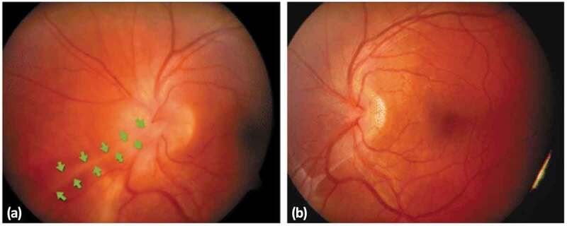
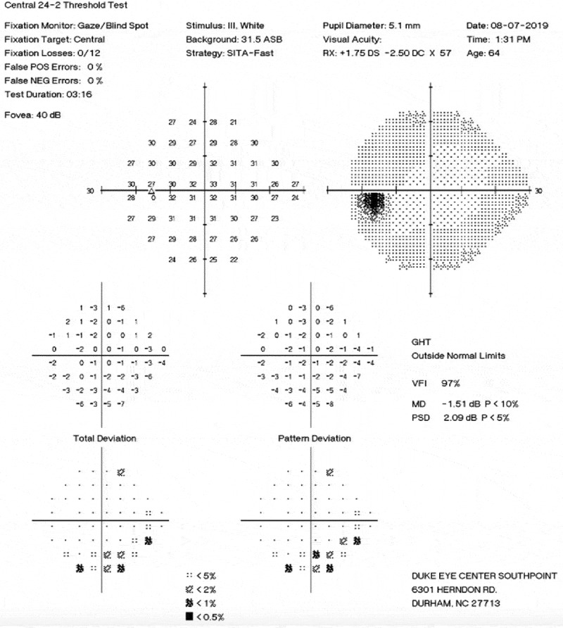
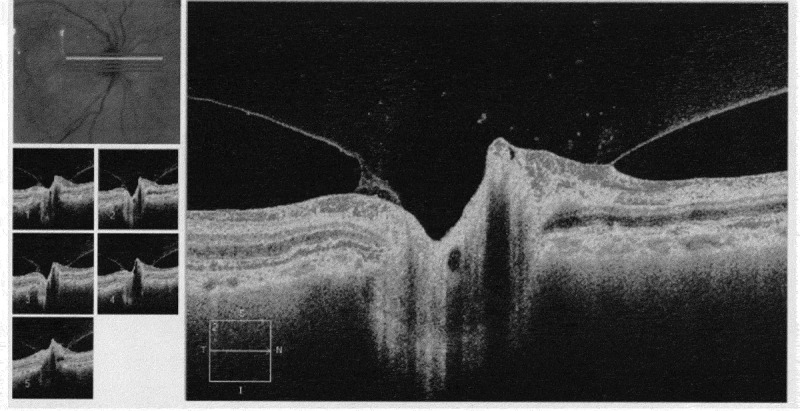
Vitreopapillary traction (VPT) syndrome is a potentially visually significant disorder of the vitreopapillary interface characterised by an incomplete posterior vitreous detachment with the persistently adherent vitreous exerting tractional pull on the optic disc and resulting in morphologic alterations and a consequent decline of visual function. It is most commonly unilateral but bilateral reports have also been described. The cause of the condition may be unknown or idiopathic, although the histology of traction shows proliferation of fibrous astrocytes, myofibroblasts, fibrocytes, and retinal pigment epithelial cells. It is theorised that VPT may induce a congested optic disc with neuronal dysfunction as well as decreased prelaminar flow. The present study reviews and summarises the features, diagnosis, and management of VPT.

Keywords: Vitreopapillary traction; optic disc; optic neuropathy; posterior vitreous detachment; visual field loss.


Figures

Figure 1.
Figure 1.
Fundus photographs demonstrating vitreopapillary traction. (a) Photographs of the left eye showing adhesions extending beyond the disc. (b) Postoperative photo showing improvement in retinal vasculature and disc margins.
Figure 2.
Figure 2.
A 24–2 visual field of a patient who presented with the complaint of a “black kidney bean smudge” in her vision. The patient was found to have a VPT.
Figure 3.
Figure 3.
Optical coherence tomography showing various horizontal slices of the vitreopapillary traction centred on the optic nerve.

Similar articles

Cited by

References

    1. Moeschlin Sandoz Y, Monatsbl K, Augenh F.. 108:310, 1942 (Cited by).//Schepens CL. Clinical aspects of pathologic changes in the vitreous body. Am J Ophthalmol. 1954;38(no.l,pt.2):8–21. doi:10.1016/0002-9394(54)90004-5. - DOI - PubMed
    1. Katz B, Hoyt WF. Intrapapillary and peripapillary hemorrhage in young patients with incomplete posterior vitreous detachment. Signs of vitreopapillary traction. Ophthalmology. 1995. February;102(2):349–354. PubMed PMID: 7862424. doi:10.1016/S0161-6420(95)31018-4. - DOI - PubMed
    1. Los LI, van der Worp RJ, van Luyn MJA, Hooymans JMM. Age-related liquefaction of the human vitreous body: LM and TEM evaluation of the role of proteoglycans and collagen. Invest Ophthalmol Visual Sci. 2003;44(7):2828. doi:10.1167/iovs.02-0588. - DOI - PubMed
    1. Ito Y, Terasaki H, Suzuki T, et al. Mapping posterior vitreous detachment by optical coherence tomography in eyes with idiopathic macular hole. Am J Ophthalmol. 2003;135(3):351–355. doi:10.1016/s0002-9394(02)01944-x. - DOI - PubMed
    1. Stalmans P, Duker JS, Kaiser PK, et al. OCT-based interpretation of the vitreomacular interface and indications for pharmacologic vitreolysis. Retina. 2013;33(10):2003–2011. doi:10.1097/iae.0b013e3182993ef8. - DOI - PubMed