Case Reports
. 2019 Feb 26;44(5):319-326.
doi: 10.1080/01658107.2019.1583761.

Neuro-Ophthalmic Sarcoidosis

Affiliations
Case Reports

Neuro-Ophthalmic Sarcoidosis

Giovanni Campagna et al. Neuroophthalmology. .

Abstract

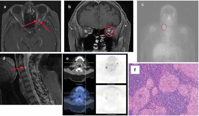
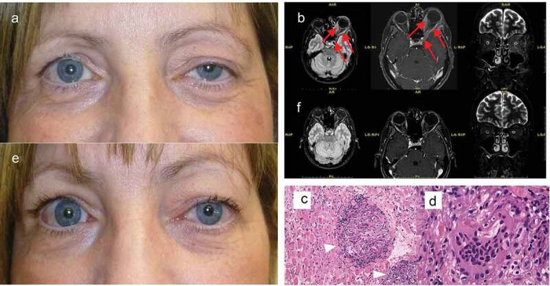
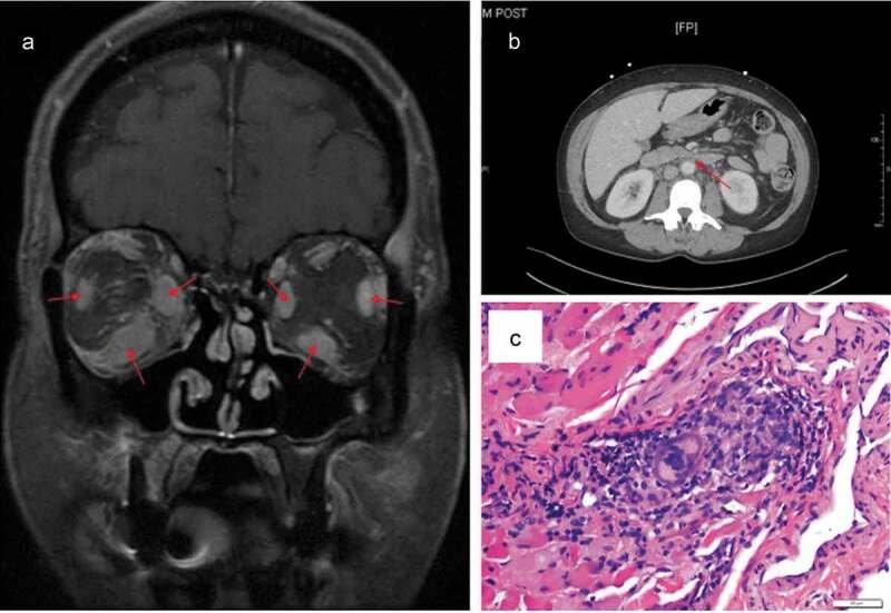
Background: In the absence of confirmatory biopsy, the criteria for diagnosis of neuro-ophthalmic sarcoidosis are not well established. Diagnostic criteria for both intraocular sarcoidosis and neurosarcoidosis have been proposed, but the diagnosis of neuro-ophthalmic sarcoidosis remains challenging. It is our intention to augment what is currently known about the diagnosis of neuro-ophthalmic sarcoidosis by providing a series of biopsy-proven cases that contribute to the continued development of diagnostic criteria for this enigmatic condition. Methods: Case series of four Caucasian women with biopsy-proven neuro-ophthalmic sarcoidosis. Results: The first patient was initially diagnosed with traumatic optic neuropathy following a fall. Years later, the presence of pathologic submandibular lymphadenopathy was identified and biopsied, revealing non-caseating granulomas. The second and third cases involved sarcoidosis of the extraocular muscles without clear or common systemic features of sarcoidosis. In the fourth and final case, the patient presented with a Horner syndrome attributed to sarcoid infiltration of the ipsilateral sympathetic chain. Bronchoscopy with biopsy showed non-caseating granulomas consistent with sarcoidosis. Conclusions: We describe four cases of neuro-ophthalmic sarcoidosis and propose possible neuro-orbital and neuro-ophthalmic criteria both with and without diagnostic biopsy.

Keywords: Neuro-ophthalmic sarcoidosis; diagnostic criteria; non-caseating granuloma; sarcoidosis.


Figures

Figure 1.
Figure 1.
Case 1. (a) Axial T1 MRI showing mid-orbital thickening and enhancement of the left optic nerve sheath with a “tram-tracking” appearance. (b) Coronal T1 MRI at the posterior orbital apex showing soft-tissue enhancing mass surrounding the left optic nerve. (c) Gallium scan showing lymphadenopathy in the right cervical parajugular level III region. (d) Spinal sagittal T1 MRI showing an enhancing nodule at the level of C3. (e) PET scan showing bilateral neck lymphadenopathy. (f) Histological biopsy specimen with Hematoxylin-eosin staining of the left submandibular lymph node showing non-caseating granulomatous inflammation with focal necrosis and hyalinisation.
Figure 2.
Figure 2.
Case 2. (a) External appearance at presentation notable for left proptosis and upper eyelid ptosis. (b) After three months of steroid treatment the ptosis reduced. Brain MRI (Left: axial FLAIR; Middle: axial T1-weighted; Right: coronal long TE). (c) At presentation there is involvement of EOMs and tendons associated with a 3 mm proptosis of the left eye. (d) Three months later, after steroid treatment, there is improvement of proptosis and EOMs have returned to normal limits. Hematoxylin-eosin staining of the medial rectus muscle biopsy. (e) Multiple non-caseating sarcoid-type granulomas (arrowheads; magnification 20x) suggestive of sarcoidosis. (f) Higher magnification (63x) of a multinucleated giant cell.
Figure 3.
Figure 3.
Case 3. (a) T1 fat-saturated coronal MRI of the orbit shows enlarged muscle bellies inferiorly, medially, and laterally in both eyes. The orbital fat and septum are enhancing. (b) CT of the abdomen reveals diffuse retroperitoneal fibrosis encasing the coeliac trunk, superior mesenteric artery, and right renal artery. (c) Hematoxylin-eosin stain of specimen obtained from the right lateral rectus showing infiltration of histiocytes, lymphocytes, and giant cells in skeletal muscle.

References

    1. Stern BJ, Aksamit A, Clifford D, Scott TF.. Neurologic presentations of neurosarcoidosis. Neurol Clin. 2010;28(1):185–198. doi:10.1016/j.ncl.2009.09.012. - DOI - PubMed
    1. Allen RKA, Stellars RE, Sandstrom PA. A prospective study of 32 patients with neurosarcoidosis. Sarcoidosis Vasc Diffuse Lung Dis. 2003;20:118–215. - PubMed
    1. Lacomis D. Neurosarcoidosis. Curr Neuropharmacol. 2011;9(3):429–436. doi:10.2174/157015911796557975. - DOI - PMC - PubMed
    1. Jabs DA, Johns CJ. Ocular involvement in chronic sarcoidosis. Am J Ophthalmol. 1986;102(3):297–301. doi:10.1016/0002-9394(86)90001-2. - DOI - PubMed
    1. Braswell RA, Kline LB. Neuro-ophthalmologic manifestations of sarcoidosis. Int Ophthalmol Clin. 2007;47:67–77. doi:10.1097/IIO.0b013e3181571d65. - DOI - PubMed

Publication types

LinkOut - more resources