Review
. 2020 Sep 8:13:581097.
doi: 10.3389/fnmol.2020.581097. eCollection 2020.

The Structure, Function, and Physiology of the Fetal and Adult Acetylcholine Receptor in Muscle

Affiliations
Review

The Structure, Function, and Physiology of the Fetal and Adult Acetylcholine Receptor in Muscle

Hakan Cetin et al. Front Mol Neurosci. .

Abstract

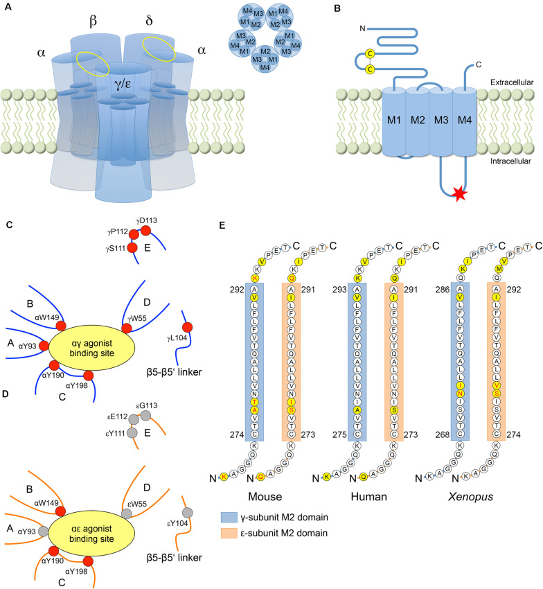
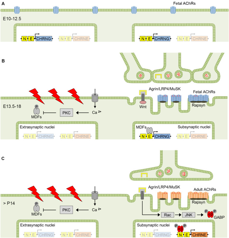
The neuromuscular junction (NMJ) is a highly developed synapse linking motor neuron activity with muscle contraction. A complex of molecular cascades together with the specialized NMJ architecture ensures that each action potential arriving at the motor nerve terminal is translated into an action potential in the muscle fiber. The muscle-type nicotinic acetylcholine receptor (AChR) is a key molecular component located at the postsynaptic muscle membrane responsible for the generation of the endplate potential (EPP), which usually exceeds the threshold potential necessary to activate voltage-gated sodium channels and triggers a muscle action potential. Two AChR isoforms are found in mammalian muscle. The fetal isoform is present in prenatal stages and is involved in the development of the neuromuscular system whereas the adult isoform prevails thereafter, except after denervation when the fetal form is re-expressed throughout the muscle. This review will summarize the structural and functional differences between the two isoforms and outline congenital and autoimmune myasthenic syndromes that involve the isoform specific AChR subunits.

Keywords: adult acetylcholine receptor; fetal acetylcholine receptor; ion channel; muscle development; myasthenia; neuromuscular junction.


Figures

FIGURE 1
FIGURE 1
Structural differences between the γ- and ε-subunit. (A) AChR consisting of five subunits with the fetal specific γ-subunit or the adult specific ε-subunit. Each subunit has four helical transmembrane domains M1-M4. The αγ/αε and αδ agonist binding sites are located between the corresponding subunits (yellow ellipses). (B) AChR subunit with N-terminal extracellular domain containing the Cys-loop, four transmembrane domains and a cytoplasmic loop domain between M3 and M4. In human, a consensus sequence for phosphorylation by protein kinase A (RRXSX, where X is any amino acid) is only present in the M3-M4 loop of the ε-subunit (red asterisk) without any corresponding sequence in the γ-subunit (residues 370–374 in the ε-subunit, i.e., RRASS). (C) At the αγ binding site, the favorable free energy resulting from the change of ACh affinities upon transformation of the AChR from the resting into the open-channel state is provided by the five aromatic core residues αY93, αW149, αY190, αY198, and γW55 from discontinuous sections of the primary sequence that rearrange close to the binding site upon agonist binding. The relative conformation of the aromatic core is stabilized by four other residues (γL104, γS111, γP112, and γD113) increasing the affinity of the αγ binding site to the agonist. (D) At the αε binding site, acetylcholine binding energy is provided by only three (instead of five) aromatic core residues (αW149, αY190, and αY198), resulting in a lower affinity to the agonist (with residues contributing to acetylcholine binding energy in red). (E) The majority of the amino acid residues of the pore forming M2 domain is highly conserved across paralogs and species. The γ- and ε-subunits differ at only three amino acid residues in mouse and Xenopus, and at two residues in human (yellow circles). The AChR isoform specific conductance was shown to be determined by amino acid residues within the M2 domain (in mouse and Xenopus) and both flanking regions (in mouse) (amino acids in red).
FIGURE 2
FIGURE 2
Factors determining fetal and adult AChR expression at the NMJ. Before innervation, fetal AChRs are evenly distributed at the surface of mouse myotubes (A), but aggregate to clusters at the myotube center between E13.5 and E18 (i.e., prepatterning), when myotubes start to become innervated (B). The exact mechanism of prepatterning is unclear but was suggested to be nerve-independent and require MuSK, LRP4 and Wnt signaling. The first 2 weeks after birth are dominated by synapse elimination resulting in singly innervated muscle fibers, and electrical activation and nerve-released agrin determine the conversion from fetal to adult AChRs, with the latter predominantly expressed in the adult muscle (C). AChR, acetylcholine receptor; GABP, GA-binding protein; JNK, c-Jun NH2-terminal kinase; MDF, myogenic determination factor; MuSK, muscle-specific kinase; LRP4, low-density lipoprotein related protein 4; PKC, protein kinase C.

Similar articles

Cited by

References

    1. Adams D. J., Dwyer T. M., Hille B. (1980). The permeability of endplate channels to monovalent and divalent metal cations. J. Gen. Physiol. 75 493–510. 10.1085/jgp.75.5.493 - DOI - PMC - PubMed
    1. Allen N. M., Hacohen Y., Palace J., Beeson D., Vincent A., Jungbluth H. (2016). Salbutamol-responsive fetal acetylcholine receptor inactivation syndrome. Neurology 86 692–694. 10.1212/wnl.0000000000002382 - DOI - PMC - PubMed
    1. Asher O., Provenzano C., Fuchs S. (1991). Regulation of acetylcholine receptor gene expression in rats treated with alpha-bungarotoxin. FEBS Lett. 282 242–246. 10.1016/0014-5793(91)80487-n - DOI - PubMed
    1. Auerbach A., Akk G. (1998). Desensitization of mouse nicotinic acetylcholine receptor channels. A two-gate mechanism. J. Gen. Physiol. 112 181–197. 10.1085/jgp.112.2.181 - DOI - PMC - PubMed
    1. Barnes P. R., Kanabar D. J., Brueton L., Newsom-Davis J., Huson S. M., Mann N. P., et al. (1995). Recurrent congenital arthrogryposis leading to a diagnosis of myasthenia gravis in an initially asymptomatic mother. Neuromuscul. Disord. 5 59–65. 10.1016/0960-8966(94)e0029-8 - DOI - PubMed

LinkOut - more resources