. 2020 Sep 4:11:1322.
doi: 10.3389/fphar.2020.01322. eCollection 2020.

Unraveling the Pharmacological Potential of Lichen Extracts in the Context of Cancer and Inflammation With a Broad Screening Approach

Affiliations

Unraveling the Pharmacological Potential of Lichen Extracts in the Context of Cancer and Inflammation With a Broad Screening Approach

Rebecca Ingelfinger et al. Front Pharmacol. .

Abstract

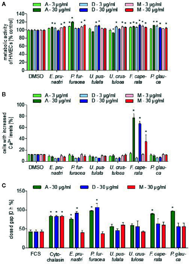
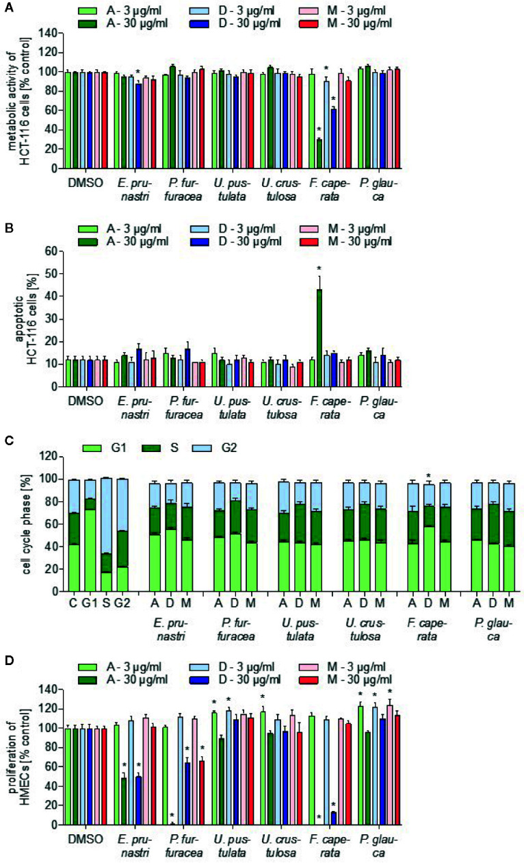
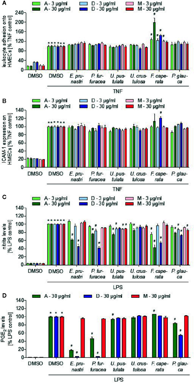
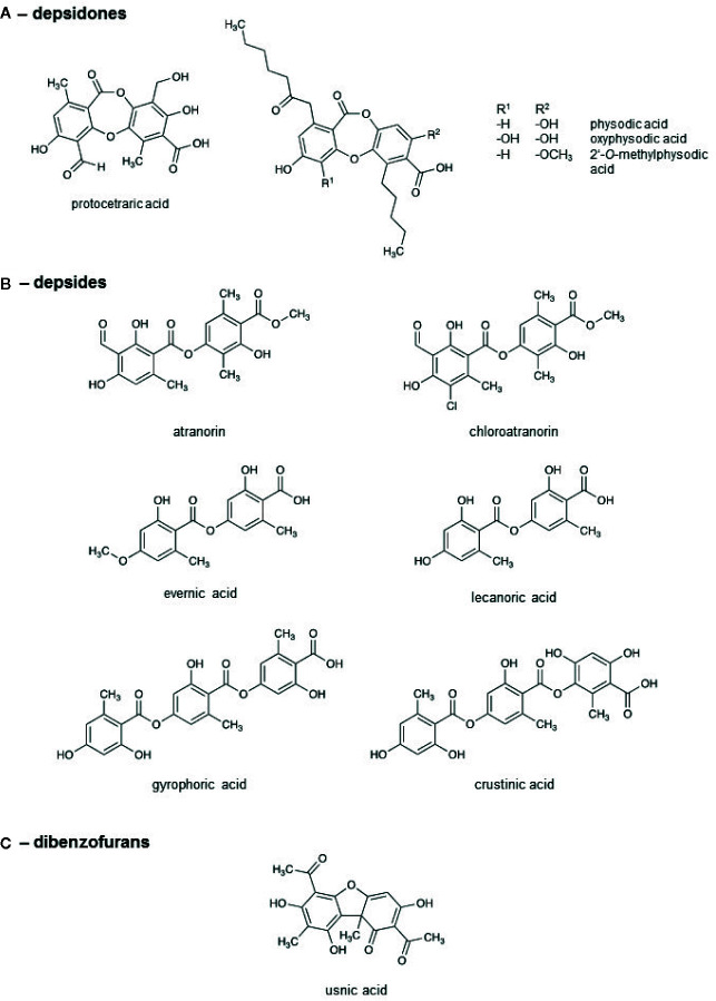
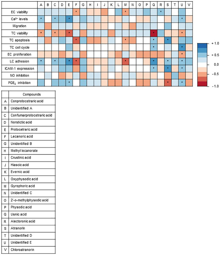
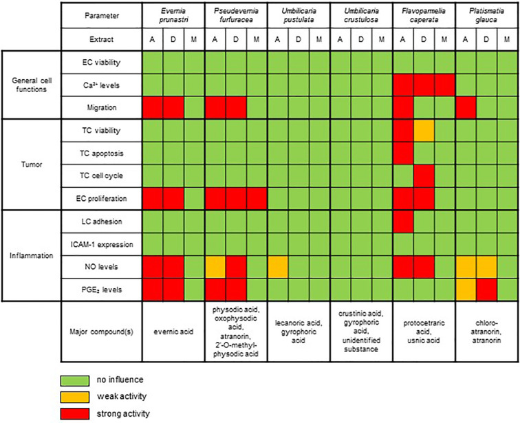
Lichen-forming fungi are symbiotic organisms that synthesize unique natural products with potential for new drug leads. Here, we explored the pharmacological activity of six lichen extracts (Evernia prunastri, Pseudevernia furfuracea, Umbilicaria pustulata, Umbilicaria crustulosa, Flavoparmelia caperata, Platismatia glauca) in the context of cancer and inflammation using a comprehensive set of 11 functional and biochemical in vitro screening assays. We assayed intracellular Ca2+ levels and cell migration. For cancer, we measured tumor cell proliferation, cell cycle distribution and apoptosis, as well as the angiogenesis-associated proliferation of endothelial cells (ECs). Targeting inflammation, we assayed leukocyte adhesion onto ECs, EC adhesion molecule expression, as well as nitric oxide production and prostaglandin (PG)E2 synthesis in leukocytes. Remarkably, none of the lichen extracts showed any detrimental influence on the viability of ECs. We showed for the first time that extracts of F. caperata induce Ca2+ signaling. Furthermore, extracts from E. prunastri, P. furfuracea, F. caperata, and P. glauca reduced cell migration. Interestingly, F. caperata extracts strongly decreased tumor cell survival. The proliferation of ECs was significantly reduced by E. prunastri, P. furfuracea, and F. caperata extracts. The extracts did not inhibit the activity of inflammatory processes in ECs. However, the pro-inflammatory activation of leukocytes was inhibited by extracts from E. prunastri, P. furfuracea, F. caperata, and P. glauca. After revealing the potential biological activities of lichen extracts by an array of screening tests, a correlation analysis was performed to evaluate particular roles of abundant lichen secondary metabolites, such as atranorin, physodic acid, and protocetraric acid as well as usnic acid in various combinations. Overall, some of the lichen extracts tested in this study exhibit significant pharmacological activity in the context of inflammation and/or cancer, indicating that the group lichen-forming fungi includes promising members for further testing.

Keywords: cancer; cytotoxicity; inflammation; lichen extracts; migration; screening.


Figures

Figure 1
Figure 1
Effects of lichen extracts on general cell functions. (A) For the cell viability assay, HMEC-1 cells were grown to confluence and treated with lichen extracts (3 and 30 µg/ml) or dimethyl sulfoxide (DMSO) (control) for 24 h. Resazurin-containing CellTiter-Blue reagent was added for the last 4 h of treatment. The metabolic activity was quantified by fluorescence measurements of resorufin. Data are expressed as mean ± SEM. n=2, *p ≤ 0.05 versus control. (B) For the Ca2+-assay, HEK293 cells were preincubated with Fluo-8-AM. Lichen extracts (3 and 30 µg/ml) or DMSO (control) were added for 5 min. Data are expressed as mean ± SEM. n=3, *p ≤ 0.05 versus control. (C) For the scratch assay, the NIH3T3 monolayer was scraped in a straight line and thereafter treated with 30 µg/ml of lichen extracts or with 650 nM cytochalasin (positive control) or DMSO (control) for 24 h. The size of the gap after 12.5 h was related to the size of the gap at 0 h and shown as % value. Data are expressed as mean ± SEM. n=2, *p ≤ 0.05 versus control.
Figure 2
Figure 2
Toxicity characterization of lichen extracts. (A) For the cell viability assay, HCT-116 cells were incubated with 3 or 30 µg/ml lichen extracts or dimethyl sulfoxide (DMSO) (control) for 24 h. WST-1 dye was added for 60 min and the viability was quantified by detection of formazan. Data are expressed as mean ± SEM. n=2, *p ≤ 0.05 versus control. (B) For the apoptosis assay, HCT-116 cells were incubated with 3 and 30 µg/ml lichen extracts or DMSO (control) for 24 h. Apoptotic cells were stained with a caspase-3-detecting antibody. Data are expressed as mean ± SEM. n=2, *p ≤ 0.05 versus control. (C) For the cell cycle analysis, HCT-116 cells were incubated for 24 h with 30 µg/ml lichen extracts or DMSO (control). Cells were stained with propidium iodide and measured using flow cytometry. Data are expressed as mean ± SEM. n=3, *p ≤ 0.05 versus control. Significances are shown when all three cell cycle phases were significantly different. (D) For the proliferation assay, HMEC-1 cells were grown in low-density and treated after 24 h with lichen extracts (3 and 30 µg/ml) for 72 h. Cells were stained with crystal violet solution. The amount of DNA-bound crystal violet was detected by absorbance measurements. Data are expressed as mean ± SEM. n=2, *p ≤ 0.05 versus control.
Figure 3
Figure 3
Effects of lichen extracts on cellular functions. (A, B) HMEC-1 cells were grown to confluence, preincubated with lichen extracts (3 and 30 µg/ml) for 30 min, and activated with TNF (10 ng/ml) for 24 h. (A) For the leukocyte adhesion assay, untreated THP-1 cells were stained with CellTracker Green and were allowed to adhere onto the treated endothelial cells (ECs) for 5 min. Non-adherent THP-1 cells were removed by washing. The adhesion of leukocytes onto ECs was quantified by fluorescence measurements. (B) For ICAM-1 expression analysis, cells were incubated with a fluorescein isothiocyanate (FITC)-labeled ICAM-1 antibody for 45 min and the cell surface expression of ICAM-1 was quantified by fluorescence measurements after washing. (A, B) Data are expressed as mean ± SEM. n=2, *p ≤ 0.05 versus dimethyl sulfoxide (DMSO) control; #p ≤ 0.05 TNF control. (C, D) For the nitric oxide (NO) (C) and prostaglandin E2 (PGE2) (D) inhibition assay, RAW macrophages were preincubated with 3 or 30 µg/ml lichen extracts or DMSO (control) for 30 min followed by the addition of 100 ng/ml lipopolysaccharide (LPS) for 24 h. Data are expressed as mean ± SEM. n=3, *p ≤ 0.05 versus DMSO control; #p ≤ 0.05 LPS control.
Figure 4
Figure 4
Chemical structures of the lichen compounds.
Figure 5
Figure 5
Graphical representation of Spearman’s correlation matrix. The natural compounds detected via high performance liquid chromatography (HPLC) in the lichen extracts were correlated with the outcomes of the 11 functional and biochemical test systems. The heat map shows Spearman’s correlation between the outcomes of the test systems and the natural compounds of the lichens identified by HPLC. Each column represents an individual compound and each row defines an individual test system. Positive correlation values are in red, and negative correlation values are in blue. The correlation analysis of lichen substances and assay outcomes was conducted with R (V3.6.1 available online https://www.R-project.org/) (Macdonald et al., 1999) and visualized as correlation matrix (V0.84 available online https://github.com/taiyun/corrplot; ColorBrewer Palettes V1.1-2 available online https://CRAN.R-project.org/package=RColorBrewer). *p ≤ 0.05.
Figure 6
Figure 6
Summary of the effects of the UV-absorbing lichen extracts in the various biochemical and functional test systems.

References

    1. Ades E. W., Candal F. J., Swerlick R. A., George V. G., Summers S., Bosse D. C., et al. (1992). HMEC-1: establishment of an immortalized human microvascular endothelial cell line. J. Invest. Dermatol. 99 (6), 683–690. 10.1111/1523-1747.ep12613748 - DOI - PubMed
    1. Asplund J., Bokhorst S., Kardol P., Wardle D. A. (2015). Removal of secondary compounds increases invertebrate abundance in lichens. Fungal Ecol. 18, 18–25. 10.1016/j.funeco.2015.07.009 - DOI
    1. Backorova M., Jendzelovsky R., Kello M., Backor M., Mikes J., Fedorocko P. (2012). Lichen secondary metabolites are responsible for induction of apoptosis in HT-29 and A2780 human cancer cell lines. Toxicol. Vitro 26 (3), 462–468. 10.1016/j.tiv.2012.01.017 - DOI - PubMed
    1. Bauer J., Waltenberger B., Noha S. M., Schuster D., Rollinger J. M., Boustie J., et al. (2012). Discovery of depsides and depsidones from lichen as potent inhibitors of microsomal prostaglandin E2 synthase-1 using pharmacophore models. ChemMedChem 7 (12), 2077–2081. 10.1002/cmdc.201200345 - DOI - PMC - PubMed
    1. Benatti M. N., Gernert M., Schmitt I. (2013). Parmotrema hydrium, a new species of Parmeliaceae in southeastern Brazil. Acta Botanica Brasilica 27 (4), 810–814. 10.1590/S0102-33062013000400021 - DOI