. 2020 Sep 8:11:1340.
doi: 10.3389/fphar.2020.01340. eCollection 2020.

In Silico, Molecular Docking and In Vitro Antimicrobial Activity of the Major Rapeseed Seed Storage Proteins

Affiliations

In Silico, Molecular Docking and In Vitro Antimicrobial Activity of the Major Rapeseed Seed Storage Proteins

Mahmudur Rahman et al. Front Pharmacol. .

Abstract

Background: In addition to their use as an edible oil and condiment crop, mustard and rapeseed (Brassica napus L., B. juncea (L.) Czern., B. nigra (L.) W.D.J.Koch, B. rapa L. and Sinapis alba L.) have been commonly used in traditional medicine for relieving pain, coughs and treating infections. The seeds contain high amounts of oil, while the remaining by-product meal after oil extraction, about 40% of seed dry weight, has a low value despite its high protein-content (~85%). The seed storage proteins (SSP) 2S albumin-type napin and 12S globulin-type cruciferin are the two predominant proteins in the seeds and show potential for value adding to the waste stream; however, information on their biological activities is scarce. In this study, purified napin and cruciferin were tested using in silico, molecular docking, and in vitro approaches for their bioactivity as antimicrobial peptides.

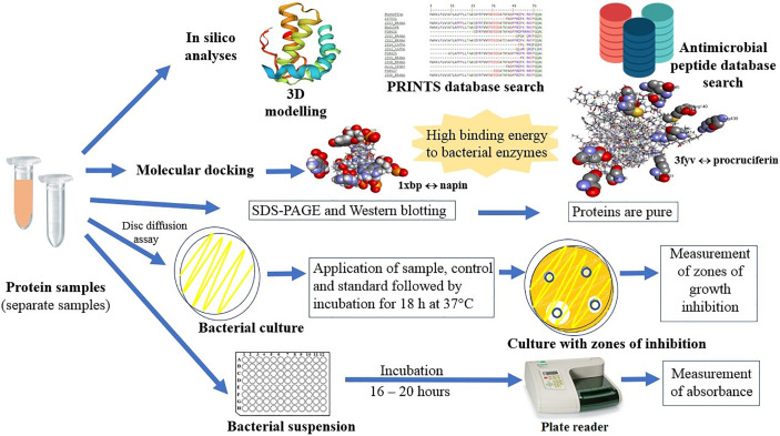
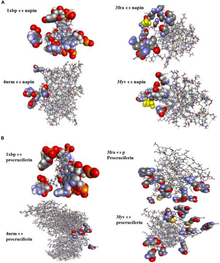
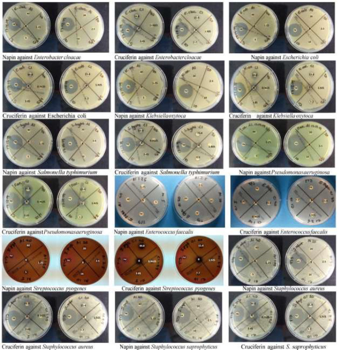
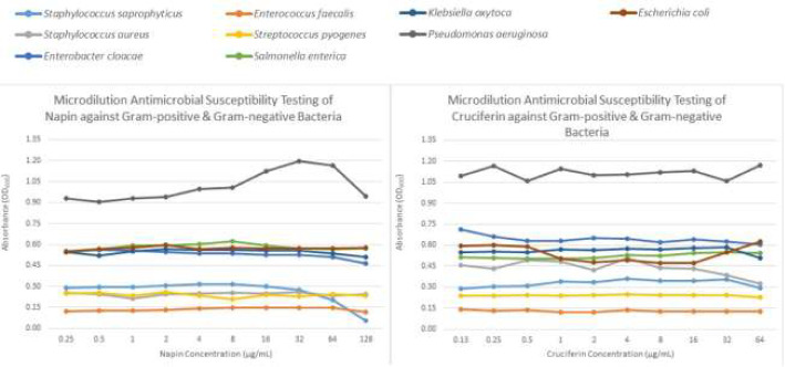
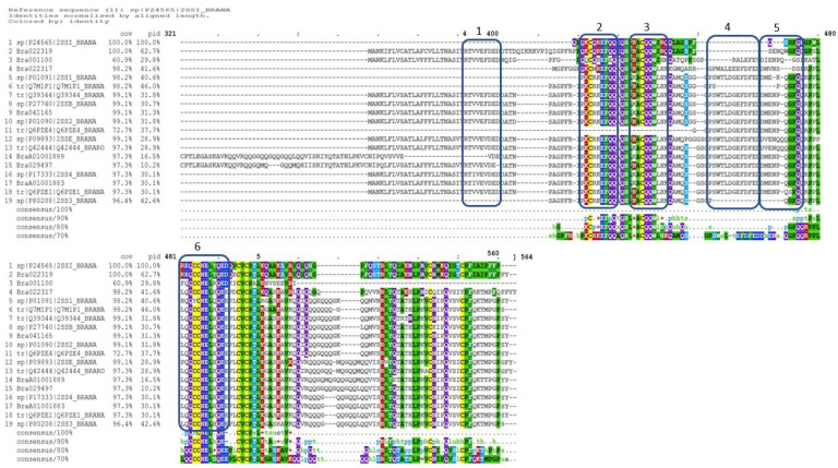
Materials and methods: The 3D-structure of 2S albumin and 12S globulin storage proteins from B. napus were investigated to predict antimicrobial activity employing an antimicrobial peptide database survey. To gain deeper insights into the potential antimicrobial activity of these SSP, in silico molecular docking was performed. The purified B. napus cruciferin and napin were then tested against both Gram-positive and Gram-negative bacteria for in vitro antimicrobial activity by disc diffusion and microdilution antimicrobial susceptibility testing.

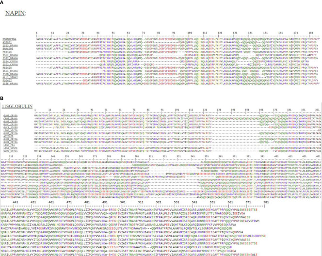
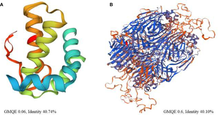
Results: In silico analysis demonstrated both SSP share similar 3D-structure with other well studied antimicrobial proteins. Molecular docking revealed that the proteins exhibited high binding energy to bacterial enzymes. Cruciferin and napin proteins appeared as a double triplet and a single doublet, respectively, following SDS-PAGE. SDS-PAGE and Western blotting also confirmed the purity of the protein samples used for assessment of antimicrobial activity. Antimicrobial susceptibility testing provided strong evidence for antimicrobial activity for the purified napin protein; however, cruciferin showed no antimicrobial activity, even at the highest dose applied.

Discussion: In silico and molecular docking results presented evidence for the potential antimicrobial activity of rapeseed cruciferin and napin SSP. However, only the in vitro antimicrobial activity of napin was confirmed. These findings warrant further investigation of this SSP protein as a potential new agent against infectious disease.

Keywords: 12S globulin; 2S albumin; cruciferin; in silico molecular docking; napin; plant antimicrobial peptide; rapeseed; seed storage protein.


Figures

Graphical Abstract
Graphical Abstract
Figure 1
Figure 1
(A) Protein motif analysis of antimicrobial signatures (shown in the same color) in napin genes using the PRINTS database. BNANAPINA = Q7DMU4_BRACM = Napin (Q7DMU4) from Brassica campestris; 2SS1_BRANA = Napin-1 (P01091), 2SSI_BRANA = Napin-1A (P24565), 2SSB_BRANA = Napin-B (P27740), 2SS2_BRANA = Napin-2 (P01090), 2SS3_BRANA = Napin-3 (P80208) and 2SSE_BRANA = Napin embryo-specific protein (P09893) are from Brassica napus, ALL1_SINAL = Allergen Sin a 1 (P09893) is from Sinapis alba, and 2SS2_CAPMA = Sweet protein mabinlin-2 (P30233) and 2SS4_CAPMA =Sweet protein mabinlin-4 (P80353) are from Capparis masaikai. The values in the parenthesis are the Uniprot accession numbers ( Supplementary Table 1 ). (B) Protein motif analysis of antimicrobial signatures (shown in the same color) in cruciferin genes using the PRINTS database. CRU3_BRANA = Cruciferin CRU1 (P33525) obtained from Brassica napus, GLUA1_ORYSJ = Glutelin type-A 1 (P07728), GLUA2_ORYSJ = Glutelin type-A 2 (P07730), GLUA3_ORYSJ = Glutelin type-A 3 (Q09151), GLUB1_ORYSJ = Glutelin type-B 1 (P14323) are obtained from Oryza sativa subsp. japonica; GLYG1_SOYBN = Glycinin G1 (P04776) from Glycine max, LEGA_GOSHI = Legumin A (P09802) from Gossypium hirsutum, LEGB6_VICFA = Legumin type B (P16079) from Vicia faba, LEGK_PEA = Legumin K (P05693) from Pisum sativum. The values in the parenthesis are the Uniprot accession numbers ( Supplementary Table 1 ).
Figure 2
Figure 2
High resolution three-dimensional theoretical structural model of (A) napin from rapeseed (Brassica napus) (P09893) based on the structure of the antibacterial sweet protein mabinlin-2 (PDB ID: 2DS2) the template protein and (B) cruciferin based on the structure of antibacterial soybean glycinin (P04776, PDB ID: 1FXZ) from Glycine max the template protein. % identity is between the target protein and the template protein.
Figure 3
Figure 3
(A) Binding interactions of napin ligand with different proteins using Patchdock. 1XBP is the 50S ribosomal subunit from Deinococcus radiodurans, 3FRA is Staphylococcus aureus dihydrofolate reductase, 4URM is S. aureus gyrase B, and 3FYV is S. aureus dihydrofolate reductase. (B) Binding interactions of procruciferin with different proteins using Patchdock. 1XBP is the 50S ribosomal subunit from Deinococcus radiodurans, 3FRA is Staphylococcus aureus dihydrofolate reductase, 4URM is S. aureus gyrase B, and 3FYV is S. aureus dihydrofolate reductase.
Figure 4
Figure 4
Plate images of disc diffusion antimicrobial susceptibility testing as per EUCAST guidelines for napin and cruciferin proteins against four Gram positive and five Gram negative bacteria, labeled electronically to reflect the actual concentrations tested. The agar plates used for the testing of the purified napin protein are labeled with the doses corresponding to 40 µg down to 1.25 µg, while plates used to test the purified cruciferin protein are labeled with the doses corresponding to 30.8 µg down to 0.9625 µg. The plates were labeled prior to the solubilization of the proteins (and subsequently, lower working concentrations were prepared for cruciferin 1, due to additional solvents added for solubilization). +v indicates that positive control antibiotics showing the expected zones of inhibition ≥14 mm (1.25 µg/23.75µg of Trimethoprim/Sulfamethoxazole) and ≥26 mm (5-µg Ciprofloxacin). B at the center indicates the blank negative control.
Figure 5
Figure 5
Graph showing the antimicrobial activity of napin and cruciferin against four Gram positive and five Gram negative bacteria, measured in microdilution antimicrobial susceptibility tests according to CLSI guidelines. The colors representing each bacterium are given at the top of the graph.
Figure 6
Figure 6
Multiple sequence alignment of Brassica napus napin proteins available in publicly open databases ( Supplementary Table 1 ) and napin proteins identified in Brassica rapa R-o-18 aligned using the program Clustal Omega [Rahman et al., 2016; Rahman et al., 2017; Rahman et al., 2018a; Rahman et al., 2020; Rahman, 2020 (in preparation)], indicates significant identity of the two proteins. Cov, sequence coverage; pid, percent identity.

References

    1. Aamir M., Singh V. K., Dubey M. K., Meena M., Kashyap S. P., Katari S. K., et al. (2018). In silico Prediction, Characterization, Molecular Docking, and Dynamic Studies on Fungal SDRs as Novel Targets for Searching Potential Fungicides Against Fusarium Wilt in Tomato. Front. Pharmacol. 9, 1–28. 10.3389/fphar.2018.01038 - DOI - PMC - PubMed
    1. Aboelsoud N. H. (2010). Herbal medicine in ancient Egypt. J. Med. Plants Res. 4, 082–086. 10.5897/JMPR09.013 - DOI
    1. Akbari A., Wu J. (2015). An integrated method of isolating napin and cruciferin from defatted canola meal. LWT-Food Sci. Technol. 64, 308–315. 10.1016/j.lwt.2015.05.046 - DOI
    1. Altschul S. F., Madden T. L., Schäffer A. A., Zhang J., Zhang Z., Miller W., et al. (1997). Gapped BLAST and PSI-BLAST: a new generation of protein database search programs. Nucleic Acids Res. 25, 3389–3402. 10.1093/nar/25.17.3389 - DOI - PMC - PubMed
    1. Amine C., Boire A., Kermarrec A., Renard D. (2019). Associative properties of rapeseed napin and pectin: Competition between liquid-liquid and liquid-solid phase separation. Food Hydrocolloids 92, 94–103. 10.1016/j.foodhyd.2019.01.026 - DOI