. 2020 Sep 8:11:544124.
doi: 10.3389/fphar.2020.544124. eCollection 2020.

Aucubin Attenuates Liver Ischemia-Reperfusion Injury by Inhibiting the HMGB1/TLR-4/NF-κB Signaling Pathway, Oxidative Stress, and Apoptosis

Affiliations

Aucubin Attenuates Liver Ischemia-Reperfusion Injury by Inhibiting the HMGB1/TLR-4/NF-κB Signaling Pathway, Oxidative Stress, and Apoptosis

Shilong Zhang et al. Front Pharmacol. .

Abstract

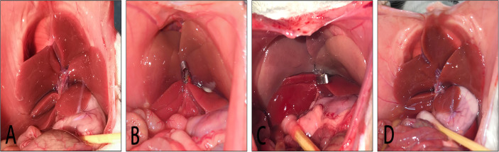
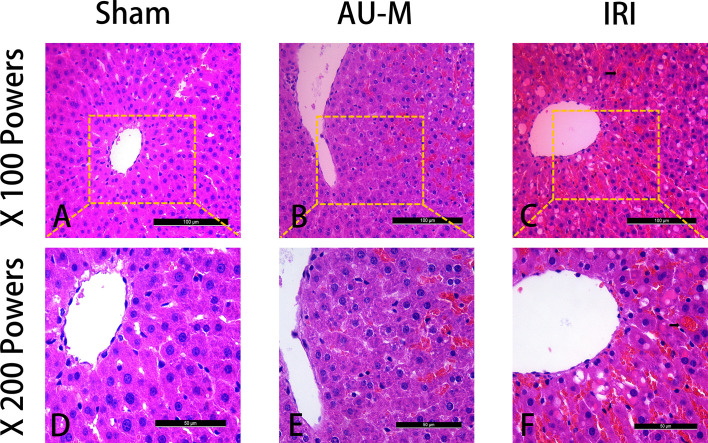
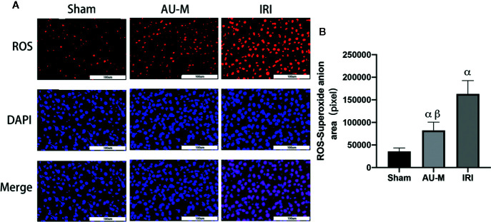
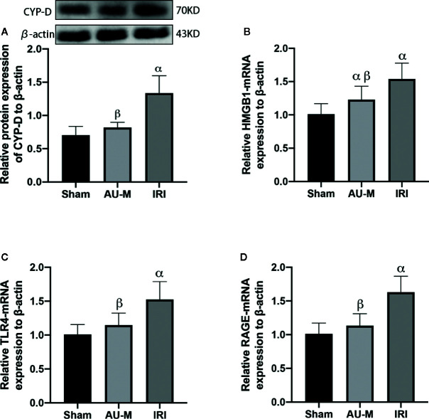
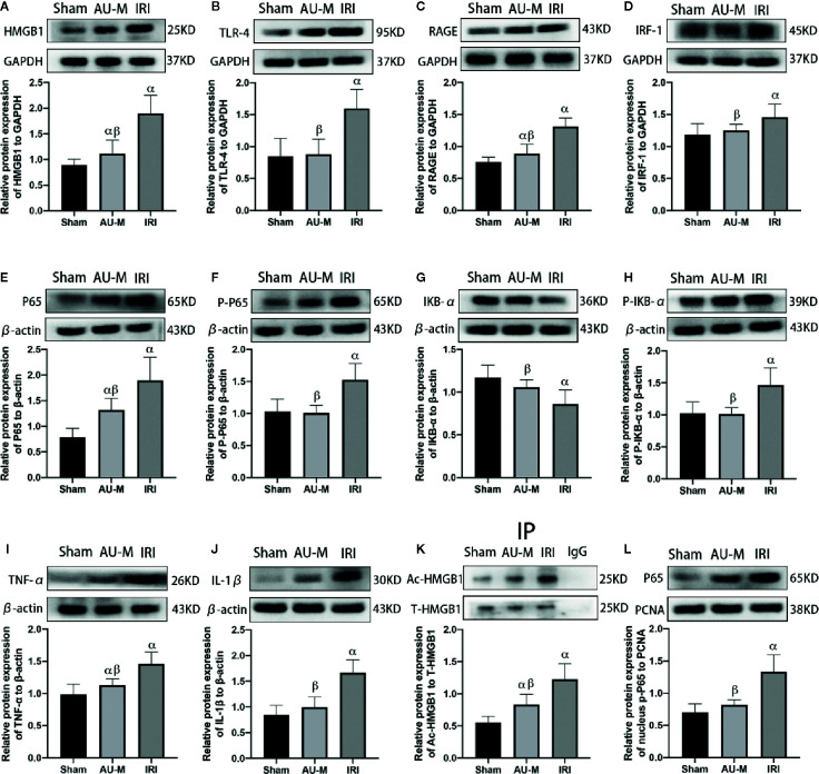
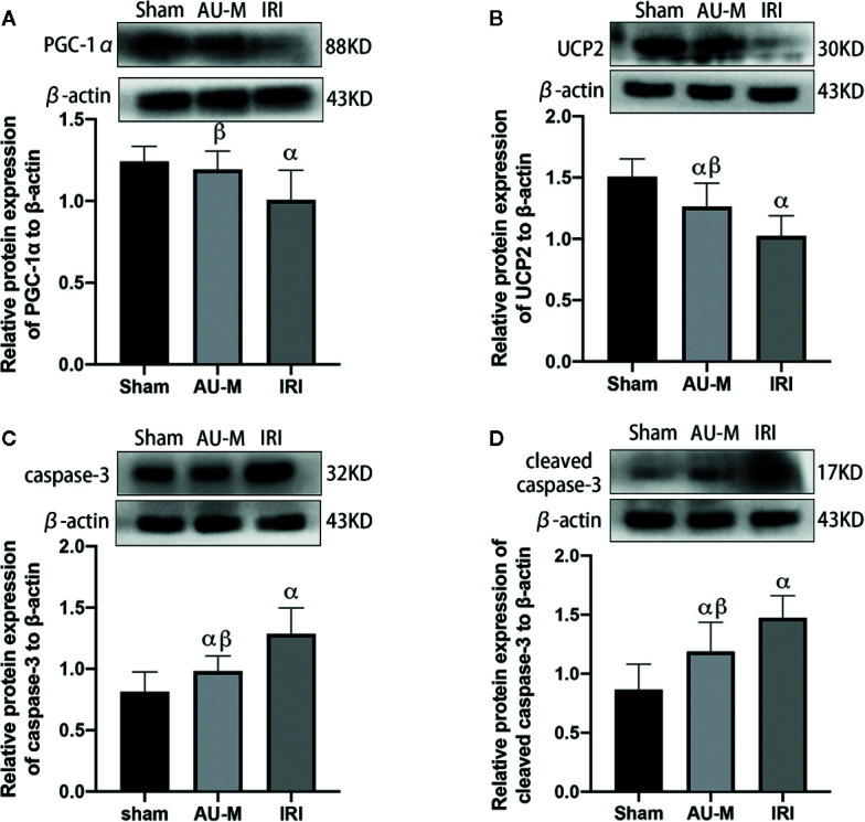
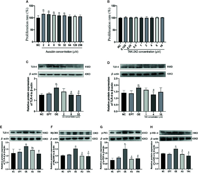
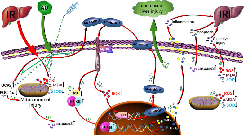
Liver ischemia-reperfusion injury (IRI) is a common clinical event with high morbidity in patients undergoing complex liver surgery or having abdominal trauma. Inflammatory and oxidative stress responses are the main contributing factors in liver IRI. The iridoid glucoside aucubin (AU) has good anti-inflammatory and antioxidative effects; however, there are no relevant reports on the protective effect of glucosides on hepatic IRI. The purpose of this study was to determine whether AU pretreatment could prevent liver IRI and to explore the mechanism. Sprague-Dawley rats were randomly divided into five groups. The sham operation and IRI control groups were given intraperitoneal injections of normal saline, while the AU low-dose (AU-L) group, AU medium-dose (AU-M) group, and AU high-dose (AU-H) group were given intraperitoneal injections of AU at doses of 1, 5, and 10 mg/kg/day, respectively. After 10 d, liver IRI (70% liver ischemia for 1 h, reperfusion for 6 h) was surgically established in all groups except the sham group. Our results confirmed that liver injury was significantly aggravated after hepatic ischemia-reperfusion. AU alleviated the increase of transaminase and pathological changes induced by ischemia-reperfusion and improved liver damage. AU could also ameliorate the inflammatory and oxidative stress responses induced by ischemia-reperfusion and reduced expression of high mobility group protein (HMG)B1, receptor for advanced glycation end-products (RAGE), tumor necrosis factor (TNF)-α, interleukin (IL)-1β, and reactive oxygen species (ROS). Moreover, AU reduced ischemia-reperfusion-induced mitochondrial dysfunction and cells apoptosis, increased peroxisome proliferator-activated receptor γ coactivator (PGC)-1α and uncoupling (UCP)2 protein expression, and reduced caspase-3, cleaved caspase-3, and Cytochrome P450 proteins (CYP) expression. To determine expression levels of the Toll-like receptor (TLR)-4/nuclear factor-κB (NF-κB) pathway-related proteins in vitro and in vivo, we also measured TLR-4, myeloid differentiation factor88 (MyD88), NF-κB P65, p-P65, I-kappa-B-alpha (IκB-α), and p-IκB-α levels. The results showed that AU effectively inhibited activation of the TLR-4/NF-κB signaling pathway. In conclusion, we showed for the first time a hepatoprotective effect for AU in liver IRI, which acted by inhibiting the HMGB1/TLR-4/NF-κB signaling pathway, oxidative stress, and apoptosis. Pretreatment with AU may be a promising strategy for preventing liver IRI.

Keywords: apoptosis; aucubin; inflammation; ischemia-reperfusion; liver; oxidative stress.


Figures

Figure 1
Figure 1
Chemical structural formula of aucubin.
Figure 2
Figure 2
Model of liver IRI. (A): liver is not ischemic; (B): liver ischemia; (C): after 1 h of liver ischemia; (D); after 6 h of liver reperfusion.
Figure 3
Figure 3
Three groups of liver sections were stained with H&E. Magnifications were low (100×) and high (200×) (scale bar=100 μm and 50 μm, respectively). The sham operation group showed normal pathology (A, D). In the IRI group, the cells showed obvious degeneration and necrosis with extensive hemorrhage (arrow) (C, F). The AU-M group showed mild degeneration and edema of the hepatocytes with occasional bleeding (B, E).
Figure 4
Figure 4
Effects of liver IR combined with intraperitoneal administration of aucubin (5 mg/kg, for 10 days) on liver ROS levels in experimental rats. Superoxide anion was expressed by red fluorescence, and nuclei by blue fluorescence; magnification was 400× and scale bar = 100 μm (A). The red fluorescence area was counted by Image Pro Plus 6.0 software analysis (B). Statistical analyses were performed using the Kolmogorov–Smirnov test followed by one-way ANOVA method. All data were expressed as mean ± SD. αSignificant difference from the sham operation group at P < 0.05. βSignificant difference from the IRI group at P < 0.05.
Figure 5
Figure 5
Effects of liver IR combined with intraperitoneal administration of aucubin (5 mg/kg, for 10 days) on protein expression of CYP-D (A) and mRNA expression of HMGB1 (B), TLR-4 (C), and RAGE (D) in the hepatic tissues of experimental rats. Statistical analyses were performed using the Kolmogorov–Smirnov test followed by One-Way ANOVA method. All data were expressed as mean ± SD. αSignificant difference from the sham operation group at P < 0.05. βSignificant difference from the IRI group at P < 0.05.
Figure 6
Figure 6
Effects of hepatic IR combined with intraperitoneal administration of aucubin (5 mg/kg, for 10 days) on quantification data and expression of HMGB1 (A), TLR-4 (B), RAGE (C), IRF-1 (D), P65 (E), p-P65 (F), IκB-α (G), p-IκB-α (H), TNF-α (I), IL-1β (J), acetylation HMGB1 (K), and intranuclear p-P65 (L), protein detected by Western blotting in the liver tissues of experimental rats. Statistical analyses were performed using the Kolmogorov–Smirnov test followed by one-way ANOVA method. All data were expressed as mean ± SD. αSignificant difference from the sham operation group at P<0.05. βSignificant difference from the IRI group at P < 0.05.
Figure 7
Figure 7
Effects of hepatic IR combined with intraperitoneal administration of aucubin (5 mg/kg, for 10 days) on quantification data and expression of PGC-1α (A), UCP2 (B), caspase-3 (C), and cleaved caspase-3 (D), protein detected by Western blotting in the liver tissues of experimental rats. Statistical analyses were performed using the Kolmogorov–Smirnov test followed by one-way ANOVA method. All data were expressed as mean ± SD. αSignificant difference from the sham operation group at P < 0.05. βSignificant difference from the IRI group at P < 0.05.
Figure 8
Figure 8
Effects of aucubin and TAK-242 on LO2 cell viability (A, B) and expression of TLR-4 (C–E), MyD88 (F), p-p65 (G), and p-IκB-α (H) proteins. Statistical analyses were performed using the Kolmogorov–Smirnov test followed by One-Way ANOVA method. All data were expressed as mean ± SD. αSignificant difference from the NC group at P < 0.05. βSignificant difference from the OE group at P <0.05. κSignificant difference from the TAK group at P < 0.05.
Figure 9
Figure 9
Aucubin attenuates liver ischemia-reperfusion injury by inhibiting the HMGB1/TLR-4/NF-κB signaling pathway, oxidative stress, and apoptosis. The molecular mechanism implicates downregulation of the HMGB1/TLR-4/NF-κB signaling pathway, oxidative stress level, mitochondrial dysfunction, and apoptosis.

Similar articles

Cited by

References

    1. Abdel-Gaber S. A., Geddawy A., Moussa R. A. (2019). The hepatoprotective effect of sitagliptin against hepatic ischemia reperfusion-induced injury in rats involves Nrf-2/HO-1 pathway. Pharmacol. Rep. 71 (6), 1044–1049. 10.1016/j.pharep.2019.06.006 - DOI - PubMed
    1. Alexandra V., Yosef K. D., Keishi N., Elizabeth A P., Cindy L., Michael E G., et al. (2020). Sleep Loss Can Cause Death through Accumulation of Reactive Oxygen Species in the Gut. Cell 181 (6), 1307–1328. 10.1016/j.cell.2020.04.049 - DOI - PubMed
    1. Alva N., Bardallo R. G., Basanta D., Palomeque J., Carbonell T. (2018). Preconditioning-Like Properties of Short-Term Hypothermia in Isolated Perfused Rat Liver (IPRL) System. Int. J. Mol. Sci. 19 (4), 1023–1035. 10.3390/ijms19041023 - DOI - PMC - PubMed
    1. Andersson U., Tracey K. J. (2011). HMGB1 is a therapeutic target for sterile inflammation and infection. Annu. Rev. Immunol. 29, 139–162. 10.1146/annurev-immunol-030409-101323 - DOI - PMC - PubMed
    1. Bi J., Zhang J., Ren Y., Du Z., Li Q., Wang Y., et al. (2019). Irisin alleviates liver ischemia-reperfusion injury by inhibiting excessive mitochondrial fission, promoting mitochondrial biogenesis and decreasing oxidative stress. Redox Biol. 20, 296–306. 10.1016/j.redox.2018.10.019 - DOI - PMC - PubMed