

Irisin Attenuates Myocardial Ischemia/Reperfusion Injury and Improves Mitochondrial Function Through AMPK Pathway in Diabetic Mice
- PMID: 33013403
- PMCID: PMC7516196
- DOI: 10.3389/fphar.2020.565160
Irisin Attenuates Myocardial Ischemia/Reperfusion Injury and Improves Mitochondrial Function Through AMPK Pathway in Diabetic Mice
Abstract
Aims: Several recent reports have shown irisin protects the heart against ischemia/reperfusion injury. However, the effect of irisin on I/R injury in diabetic mice has not been described. The present study was designed to investigate the role of irisin in myocardial ischemia-reperfusion (MI/R) injury in diabetic mice.
Methods: A mouse model of diabetes was established by feeding wild type or gene-manipulated adult male mice with a high-fat diet. All the mice received intraperitoneal injection of irisin or PBS. Thirty minutes after injection, mice were subjected to 30 min of myocardial ischemia followed by 3h (for cell apoptosis and protein determination), 24 h (for infarct size and cardiac function).
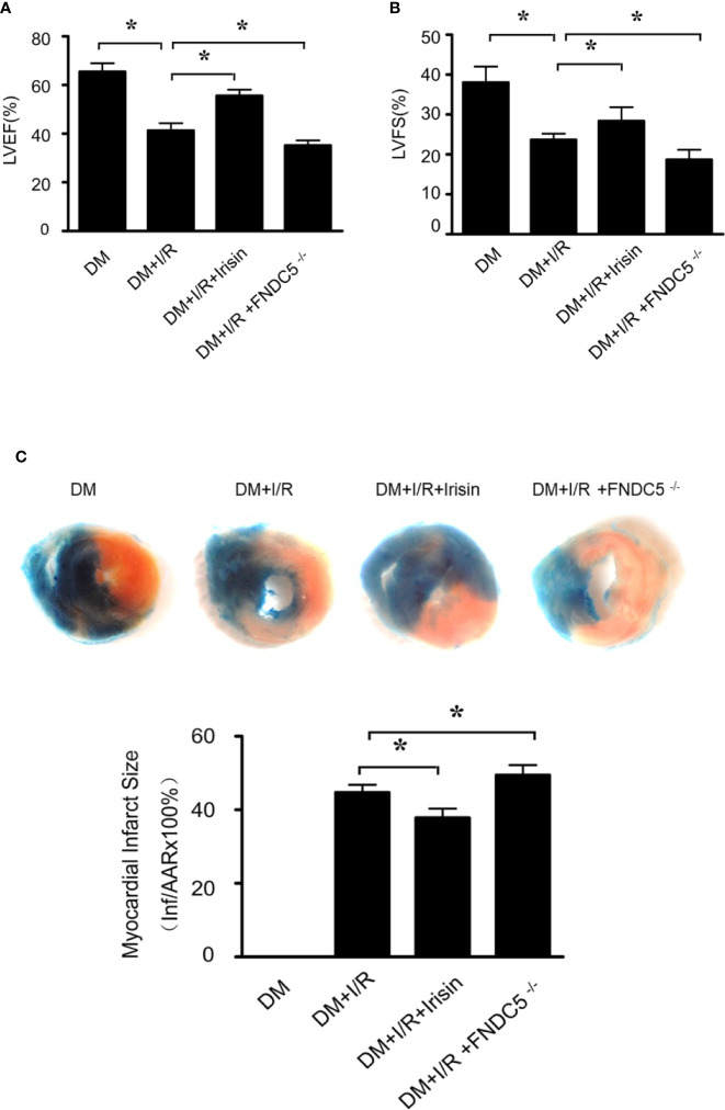
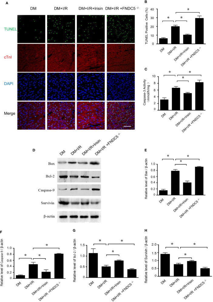
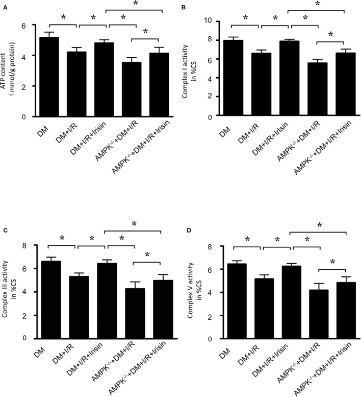
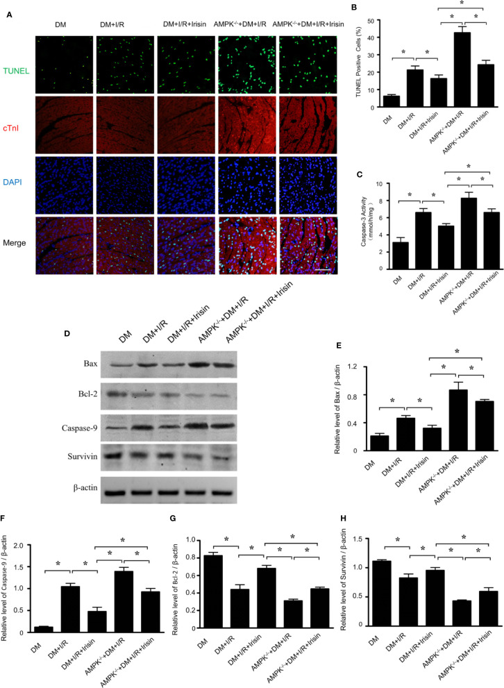
Results: Knock-out of gene FNDC5 augmented MI/R injury in diabetic mice, while irisin treatment attenuated MI/R injury, improved cardiac function, cellular ATP biogenetics, mitochondria potential, and impaired mitochondrion-related cell death. More severely impaired AMPK pathway was observed in diabetic FNDC5-/- mice received MI/R. Knock-out of gene AMPK blocks the beneficial effects of irisin on MI/R injury, cardiac function, cellular ATP biogenetics, mitochondria potential, and mitochondrion-related cell death.
Conclusions: Our present study demonstrated that irisin improves the mitochondria function and attenuates MI/R injury in diabetic mice through AMPK pathway.
Keywords: AMPK; diabetes; irisin; ischemia/reperfusion; mitochondria.
Copyright © 2020 Xin, Zhang, Gao, Ding, Yang, Wang, Liu, Guo, Yang, Zhang, Zhang, Liu, Jin and Tao.
Figures


Similar articles
-
Irisin attenuates myocardial ischemia/reperfusion-induced cardiac dysfunction by regulating ER-mitochondria interaction through a mitochondrial ubiquitin ligase-dependent mechanism.Clin Transl Med. 2020 Sep;10(5):e166. doi: 10.1002/ctm2.166. Clin Transl Med. 2020. PMID: 32997406 Free PMC article.
-
Inhibition of dynamin-related protein 1 protects against myocardial ischemia-reperfusion injury in diabetic mice.Cardiovasc Diabetol. 2017 Feb 7;16(1):19. doi: 10.1186/s12933-017-0501-2. Cardiovasc Diabetol. 2017. PMID: 28173848 Free PMC article.
-
Cardiac-derived adiponectin induced by long-term insulin treatment ameliorates myocardial ischemia/reperfusion injury in type 1 diabetic mice via AMPK signaling.Basic Res Cardiol. 2013 Jan;108(1):322. doi: 10.1007/s00395-012-0322-0. Epub 2012 Dec 22. Basic Res Cardiol. 2013. PMID: 23262803
-
FNDC5/Irisin attenuates diabetic cardiomyopathy in a type 2 diabetes mouse model by activation of integrin αV/β5-AKT signaling and reduction of oxidative/nitrosative stress.J Mol Cell Cardiol. 2021 Nov;160:27-41. doi: 10.1016/j.yjmcc.2021.06.013. Epub 2021 Jul 3. J Mol Cell Cardiol. 2021. PMID: 34224725
-
Irisin is an Effector Molecule in Exercise Rehabilitation Following Myocardial Infarction (Review).Front Physiol. 2022 Jun 29;13:935772. doi: 10.3389/fphys.2022.935772. eCollection 2022. Front Physiol. 2022. PMID: 35845994 Free PMC article. Review.
Cited by
-
The Effect of miR-505-5p on Inhibition of Serum Uromodulin Ameliorates Myocardial Inflammation and Apoptosis Induced by Ischemia-Reperfusion.Oxid Med Cell Longev. 2022 Oct 3;2022:3521971. doi: 10.1155/2022/3521971. eCollection 2022. Oxid Med Cell Longev. 2022. PMID: 36225178 Free PMC article.
-
The Controversial Role of Irisin in Clinical Management of Coronary Heart Disease.Front Endocrinol (Lausanne). 2021 Jul 1;12:678309. doi: 10.3389/fendo.2021.678309. eCollection 2021. Front Endocrinol (Lausanne). 2021. PMID: 34276559 Free PMC article. Review.
-
Irisin and Incretin Hormones: Similarities, Differences, and Implications in Type 2 Diabetes and Obesity.Biomolecules. 2021 Feb 15;11(2):286. doi: 10.3390/biom11020286. Biomolecules. 2021. PMID: 33671882 Free PMC article. Review.
-
Ginkgolide B Protects Against Ischemic Stroke via Targeting AMPK/PINK1.Front Pharmacol. 2022 Jun 28;13:941094. doi: 10.3389/fphar.2022.941094. eCollection 2022. Front Pharmacol. 2022. PMID: 35837278 Free PMC article.
-
Exercise ameliorating myocardial injury in type 2 diabetic rats by inhibiting excessive mitochondrial fission involving increased irisin expression and AMP-activated protein kinase phosphorylation.J Diabetes. 2024 Jan;16(1):e13475. doi: 10.1111/1753-0407.13475. Epub 2023 Sep 18. J Diabetes. 2024. PMID: 37721125 Free PMC article.
References
-
- Badalzadeh R., Mohammadi M., Najafi M., Ahmadiasl N., Farajnia S., Ebrahimi H. (2012). The additive effects of ischemic postconditioning and cyclosporine-A on nitric oxide activity and functions of diabetic myocardium injured by ischemia/reperfusion. J. Cardiovasc. Pharmacol. Ther. 17 (2), 181–189. 10.1177/1074248411416118 - DOI - PubMed
LinkOut - more resources
Full Text Sources