

Non-coding RNAs in Cardiac Intercellular Communication
- PMID: 33013428
- PMCID: PMC7509180
- DOI: 10.3389/fphys.2020.00738
Non-coding RNAs in Cardiac Intercellular Communication
Abstract
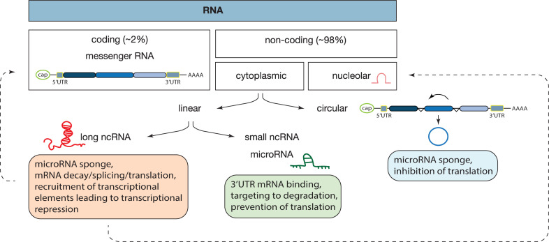
Intercellular communication allows for molecular information to be transferred from cell to cell, in order to maintain tissue or organ homeostasis. Alteration in the process due to changes, either on the vehicle or the cargo information, may contribute to pathological events, such as cardiac pathological remodeling. Extracellular vesicles (EVs), namely exosomes, are double-layer vesicles secreted by cells to mediate intercellular communication, both locally and systemically. EVs can carry different types of cargo, including non-coding RNAs (ncRNAs), which, are major regulators of physiological and pathological processes. ncRNAs transported in EVs are functionally active and trigger a cascade of processes in the recipient cells. Upon cardiac injury, exosomal ncRNAs can derive from and target different cardiac cell types to initiate cellular and molecular remodeling events such as hypertrophic growth, cardiac fibrosis, endothelial dysfunction, and inflammation, all contributing to cardiac dysfunction and, eventually, heart failure. Exosomal ncRNAs are currently accepted as crucial players in the process of cardiac pathological remodeling and alterations in their presence profile in EVs may attenuate cardiac dysfunction, suggesting that exosomal ncRNAs are potential new therapeutic targets. Here, we review the current research on the role of ncRNAs in intercellular communication, in the context of cardiac pathological remodeling.
Keywords: cardiac intercellular communication; cardiac pathological remodeling; extracellular vesicles; heart failure; non-coding RNAs.
Copyright © 2020 Videira and da Costa Martins.
Figures


Similar articles
-
Exosomal Non-Coding RNAs: Regulatory and Therapeutic Target of Hepatocellular Carcinoma.Front Oncol. 2021 Mar 26;11:653846. doi: 10.3389/fonc.2021.653846. eCollection 2021. Front Oncol. 2021. PMID: 33869059 Free PMC article. Review.
-
Extracellular Vesicles as Carriers of Non-coding RNAs in Liver Diseases.Front Pharmacol. 2018 Apr 24;9:415. doi: 10.3389/fphar.2018.00415. eCollection 2018. Front Pharmacol. 2018. PMID: 29740327 Free PMC article. Review.
-
Extracellular Non-Coding RNAs in Cardiovascular Diseases.Pharmaceutics. 2023 Jan 3;15(1):155. doi: 10.3390/pharmaceutics15010155. Pharmaceutics. 2023. PMID: 36678784 Free PMC article. Review.
-
The roles and therapeutic potential of exosomal non-coding RNAs in microglia-mediated intercellular communication.Int Immunopharmacol. 2025 Feb 20;148:114049. doi: 10.1016/j.intimp.2025.114049. Epub 2025 Jan 16. Int Immunopharmacol. 2025. PMID: 39823800 Review.
-
Extracellular Vesicle (EVs) Associated Non-Coding RNAs in Lung Cancer and Therapeutics.Int J Mol Sci. 2022 Nov 7;23(21):13637. doi: 10.3390/ijms232113637. Int J Mol Sci. 2022. PMID: 36362424 Free PMC article. Review.
Cited by
-
Serum‑derived exosomes from house dust mite‑sensitized guinea pigs contribute to inflammation in BEAS‑2B cells via the TLR4‑NF‑κB pathway.Mol Med Rep. 2021 Nov;24(5):747. doi: 10.3892/mmr.2021.12387. Epub 2021 Aug 30. Mol Med Rep. 2021. PMID: 34458929 Free PMC article.
-
Recent progress in exosomal non-coding RNAs research related to idiopathic pulmonary fibrosis.Front Genet. 2025 Mar 27;16:1556495. doi: 10.3389/fgene.2025.1556495. eCollection 2025. Front Genet. 2025. PMID: 40212286 Free PMC article. Review.
-
Extracellular Vesicles Regulate Sympatho-Excitation by Nrf2 in Heart Failure.Circ Res. 2022 Sep 30;131(8):687-700. doi: 10.1161/CIRCRESAHA.122.320916. Epub 2022 Sep 13. Circ Res. 2022. PMID: 36098045 Free PMC article.
-
Exosomal Non-coding RNA Derived from Mesenchymal Stem Cells (MSCs) in Autoimmune Diseases Progression and Therapy; an Updated Review.Cell Biochem Biophys. 2024 Dec;82(4):3091-3108. doi: 10.1007/s12013-024-01432-4. Epub 2024 Sep 3. Cell Biochem Biophys. 2024. PMID: 39225902 Review.
-
The Role of Extracellular Non-coding RNAs in Atherosclerosis.J Cardiovasc Transl Res. 2022 Jun;15(3):477-491. doi: 10.1007/s12265-022-10218-z. Epub 2022 Mar 1. J Cardiovasc Transl Res. 2022. PMID: 35233720 Review.
References
Publication types
LinkOut - more resources
Full Text Sources