Review
. 2020 Sep 9:11:1035.
doi: 10.3389/fphys.2020.01035. eCollection 2020.

HIF-1α as a Mediator of Insulin Resistance, T2DM, and Its Complications: Potential Links With Obstructive Sleep Apnea

Affiliations
Review

HIF-1α as a Mediator of Insulin Resistance, T2DM, and Its Complications: Potential Links With Obstructive Sleep Apnea

Agata Gabryelska et al. Front Physiol. .

Abstract

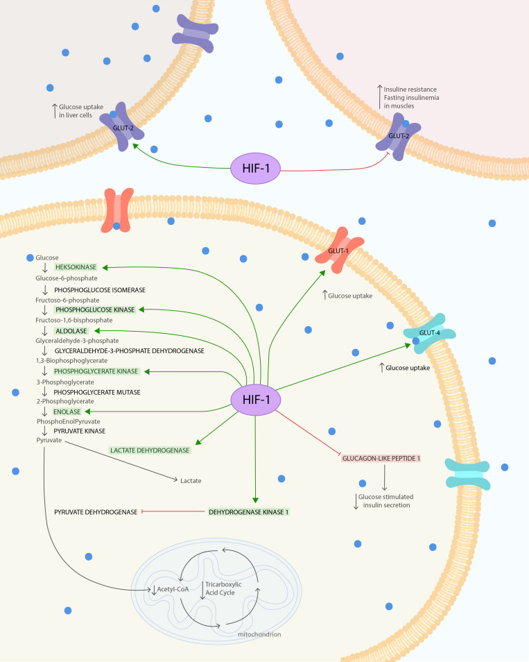
Obstructive sleep apnea syndrome (OSA) is described as an independent risk factor for the onset and progression of type 2 diabetes (T2DM), as well as for insulin resistance (IR). The mechanisms underlying these processes remain unclear. One of the proposed molecular mechanism is based on the oxygen-sensitive α-subunit of hypoxia-inducible factor 1 (HIF-1α)-a key regulator of oxygen metabolism. The concept that stabilization of HIF-1α may influence T2DM and IR is supported by cell and animal models. Cell culture studies revealed that both glucose uptake and glycolysis are regulated by HIF-1α. Furthermore, animal models indicated that increased fasting glucose may be caused by a single night with intermittent hypoxia. Moreover, in these models, hypoxia time was correlated with IR. Mice models revealed that inhibition of HIF-1α protein may downregulate fasting blood glucose and plasma insulin level. Administration of superoxide dismutase mimetic resulted in inhibition of HIF-1α protein, catecholamines, and chronic intermittent hypoxia-induced hypertension in a mice model. The hypothesis that hypoxia is an independent risk factor for IR is strengthened by experimentally confirmed improvement of insulin sensitivity among OSA patients treated with the continuous positive airway pressure. Furthermore, recent studies suggest that HIF-1α protein concentration is increased in individuals with OSA. In this literature review, we summarize the current knowledge about HIF-1α in OSA in relation to the possible pathways in which they contribute to metabolic disorders.

Keywords: HIF-1α; OSA; T2DM2; hypoxia; insulin resistance.


Figures

FIGURE 1
FIGURE 1
HIF-1α influence on glucose metabolism and insulin resistance. The HIF is composed of both oxygen-regulated a-subunit and constitutively expressed b-subunit. HIF-1α protein is highly unstable under normoxia condition. Hypoxia leads to stabilization of HIF-1α. HIF-1α under hypoxic conditions causes several changes in glucose metabolism. It increases glucose uptake via glucose transporters GLUT-1 and GLUT-4 to cells. In the cell, glucose is used in glycolysis, which is also enhanced due to the modulated expression of glycolytic enzymes: phosphoglycerate kinase-1, hexokinase 1, hexokinase 2, aldolase A, enolase 1, and phosphofructokinase L. The final product of glycolysis–pyruvate–is mostly converted to lactate instead of acetyl-CoA, due to the increased lactate dehydrogenase A activity and pyruvate decarboxylation inhibition. Pyruvate dehydrogenase kinase 1 (PDK1) action leads to suppression of dehydrogenase complex (PDH) through its phosphorylation and thereby inhibits pyruvate decarboxylation. Secondary to the decreased levels of acetyl-CoA and the action of PDK1, the TAC is downregulated. At the same time, the increased expression of HIF-1α reduces GLUT-2 phosphorylation and its expression in skeletal muscles. This leads to increased IR and fasting insulinemia after exposure to chronic hypoxia. To compensate for this metabolic imbalance, the expression of GLUT-2 in liver is increased. The glucose tolerance can also be impaired by upregulation of HIF-1α, leading to GLP-1 downregulation, which causes reduction in glucose-stimulated insulin secretion via pancreatic b-cells. The Figure 1 was prepared in Adobe Illustrator (Adobe Inc., San Jose, CA, United States).

References

    1. Alhawiti N. M., Al Mahri S., Aziz M. A., Malik S. S., Mohammad S. (2017). TXNIP in metabolic regulation: physiological role and therapeutic outlook. Curr. Drug Targets 18 1095–1103. 10.2174/1389450118666170130145514 - DOI - PMC - PubMed
    1. Anothaisintawee T., Reutrakul S., Van Cauter E., Thakkinstian A. (2016). Sleep disturbances compared to traditional risk factors for diabetes development: systematic review and meta-analysis. Sleep Med. Rev. 30 11–24. 10.1016/j.smrv.2015.10.002 - DOI - PubMed
    1. Badawi Y., Shi H. (2017). Relative contribution of prolyl hydroxylase-dependent and -independent degradation of HIF-1alpha by proteasomal pathways in cerebral ischemia. Front. Neurosci. 11:239. 10.3389/fnins.2017.00239 - DOI - PMC - PubMed
    1. Bessho R., Takiyama Y., Takiyama T., Kitsunai H., Takeda Y., Sakagami H., et al. (2019). Hypoxia-inducible factor-1α is the therapeutic target of the SGLT2 inhibitor for diabetic nephropathy. Sci. Rep. 9:14754. 10.1038/s41598-019-51343-1 - DOI - PMC - PubMed
    1. Boulton A. J., Whitehouse R. W. (2000). The Diabetic Foot. Available online at: http://www.ncbi.nlm.nih.gov/pubmed/28121117 (accessed January 12, 2020).

LinkOut - more resources