Review
. 2020 Sep 3:11:1063.
doi: 10.3389/fphys.2020.01063. eCollection 2020.

Circulating Extracellular Vesicles and Endothelial Damage in Sickle Cell Disease

Affiliations
Review

Circulating Extracellular Vesicles and Endothelial Damage in Sickle Cell Disease

Gabrielle Lapping-Carr et al. Front Physiol. .

Abstract

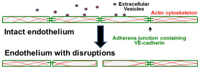
Endothelial damage is central to the pathogenesis of many of the complications of sickle cell disease. Circulating extracellular vesicles (EVs) have been implicated in modulating endothelial behavior in a variety of different, diseases with vascular pathologies. As seen in other hemolytic diseases, the plasma of sickle cell patients contains EVs of different sizes and cellular sources. The medium-sized vesicles (microparticles) primarily derive from mature red blood cells and platelets; some of these EVs have procoagulant properties, while others stimulate inflammation or endothelial adhesiveness. Most of the small EVs (including exosomes) derive from erythrocytes and erythrocyte precursors, but some also originate from platelets, white blood cells, and endothelial cells. These small EVs may alter the behavior of target cells by delivering cargo including proteins and nucleic acids. Studies in model systems implicate small EVs in promoting vaso-occlusion and disruption of endothelial integrity. Thus, both medium and small EVs may contribute to the increased endothelial damage in sickle cell disease. Development of a detailed understanding of the composition and roles of circulating EVs represents a promising approach toward novel predictive diagnostics and therapeutic approaches in sickle cell disease.

Keywords: endothelial damage; exosomes; extracellular vesicle; microvesicle; sickle cell disease.


Figures

Figure 1
Figure 1
Circulating small extracellular vesicles (EVs) from sickle cell patients cause damage to endothelial cell monolayers. (A–F) representative photomicrographs show the localization of VE-cadherin (green) and nuclei (blue in B,D,F) in endothelial cells 48 h following treatment with no EVs (A,B) or EVs from a subject with sickle cell disease (SCD) purified by precipitation (C,D) or by size exclusion chromatography (E,F). White stars indicate spaces between cells. Scale bar is 20 μm. In the examples shown, the monolayer disruption was 0% (A,B), 1.9% (C,D), or 7.5% (E,F). This figure contains different examples illustrating the observations in our previous publication (Lapping-Carr et al., 2017, 2020).
Figure 2
Figure 2
Model illustrates how circulating small EVs cause endothelial damage in sickle cell patients. In control patients or healthy patients with SCD, the endothelial cells form tight monolayers. They are held together by adhesive intercellular junctions containing VE-cadherin, and they have filamentous-actin distributed throughout the cells. In patients that develop the vasculopathy of acute chest syndrome, interaction of circulating small EVs with the endothelial cells causes disruption and loss of some adherens junctions, rearrangement of the actin cytoskeleton, and opening of spaces between cells that disrupt monolayer integrity.

Similar articles

Cited by

References

    1. Agouti I., Cointe S., Robert S., Judicone C., Loundou A., Driss F., et al. . (2015). Platelet and not erythrocyte microparticles are procoagulant in transfused thalassaemia major patients. Br. J. Haematol. 171, 615–624. 10.1111/bjh.13609, PMID: - DOI - PubMed
    1. Alaarg A., Schiffelers R. M., van Solinge W. W., van Wijk R. (2013). Red blood cell vesiculation in hereditary hemolytic anemia. Front. Physiol. 4:365. 10.3389/fphys.2013.00365, PMID: - DOI - PMC - PubMed
    1. Allan D., Limbrick A. R., Thomas P., Westerman M. P. (1981). Microvesicles from sickle erythrocytes and their relation to irreversible sickling. Br. J. Haematol. 47, 383–390. 10.1111/j.1365-2141.1981.tb02805.x, PMID: - DOI - PubMed
    1. Allan D., Limbrick A. R., Thomas P., Westerman M. P. (1982). Release of spectrin-free spicules on reoxygenation of sickled erythrocytes. Nature 295, 612–613. 10.1038/295612a0, PMID: - DOI - PubMed
    1. Almizraq R. J., Norris P. J., Inglis H., Menocha S., Wirtz M. R., Juffermans N., et al. . (2018). Blood manufacturing methods affect red blood cell product characteristics and immunomodulatory activity. Blood Adv. 2, 2296–2306. 10.1182/bloodadvances.2018021931, PMID: - DOI - PMC - PubMed