Review
. 2020 Sep 4:11:1068.
doi: 10.3389/fphys.2020.01068. eCollection 2020.

Targeting Ca2 + Handling Proteins for the Treatment of Heart Failure and Arrhythmias

Affiliations
Review

Targeting Ca2 + Handling Proteins for the Treatment of Heart Failure and Arrhythmias

Alexandra Njegic et al. Front Physiol. .

Abstract

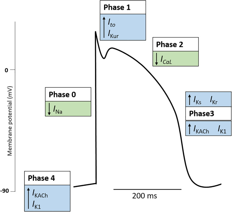
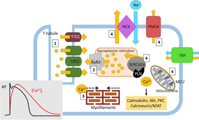
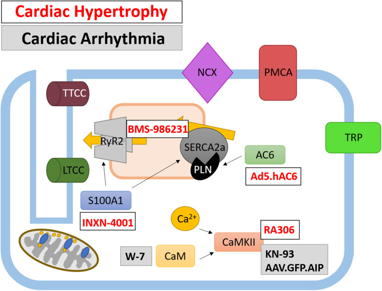
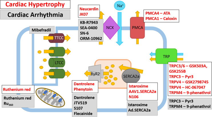
Diseases of the heart, such as heart failure and cardiac arrhythmias, are a growing socio-economic burden. Calcium (Ca2+) dysregulation is key hallmark of the failing myocardium and has long been touted as a potential therapeutic target in the treatment of a variety of cardiovascular diseases (CVD). In the heart, Ca2+ is essential for maintaining normal cardiac function through the generation of the cardiac action potential and its involvement in excitation contraction coupling. As such, the proteins which regulate Ca2+ cycling and signaling play a vital role in maintaining Ca2+ homeostasis. Changes to the expression levels and function of Ca2+-channels, pumps and associated intracellular handling proteins contribute to altered Ca2+ homeostasis in CVD. The remodeling of Ca2+-handling proteins therefore results in impaired Ca2+ cycling, Ca2+ leak from the sarcoplasmic reticulum and reduced Ca2+ clearance, all of which contributes to increased intracellular Ca2+. Currently, approved treatments for targeting Ca2+ handling dysfunction in CVD are focused on Ca2+ channel blockers. However, whilst Ca2+ channel blockers have been successful in the treatment of some arrhythmic disorders, they are not universally prescribed to heart failure patients owing to their ability to depress cardiac function. Despite the progress in CVD treatments, there remains a clear need for novel therapeutic approaches which are able to reverse pathophysiology associated with heart failure and arrhythmias. Given that heart failure and cardiac arrhythmias are closely associated with altered Ca2+ homeostasis, this review will address the molecular changes to proteins associated with both Ca2+-handling and -signaling; their potential as novel therapeutic targets will be discussed in the context of pre-clinical and, where available, clinical data.

Keywords: arrhythmia; calcium; calcium dysregulation; cardiovascular disease; heart failure.


Figures

FIGURE 1
FIGURE 1
The cardiac action potential. The cardiac action potential represents the electrical activity of a cardiomyocyte during cardiac contraction and is governed by changes in outward (blue boxes) and inward (green boxes) cardiac ion currents. Generally, the cardiac action potential is divided into five Phases. (Phase 4: Resting) The resting membrane potential is stable (∼–90 mV) and maintained by outward leak of K+ (IKACh, IK1). Here the Na+ and Ca+ channels are closed. (Phase 0: Upstroke) An action potential at a neighboring cardiomyocyte initiates a rise in membrane potential. When the membrane potential reaches the threshold potential (∼70 mV), Na+ channels open, resulting in a rapid influx of Na+ and the generation on the inward Na+ current (INa). Na+ channels become inactivated soon after opening (Phase 1: Early repolarization). Some K+ channels transiently open causing an outward K+ current (Ito, IKur). This results in a period of rapid repolarization. (Phase 2: Plateau phase) L-type Ca2+ channels are open to create a small, constant inward Ca2+ current (ICaL) which triggers excitation contraction coupling resulting in cardiomyocyte contraction. K+ currents remain active, although the two currents are electrically balanced and maintain a plateau. (Phase 3: Repolarization) Ca2+ channels become gradually inactivated and the outflow of K+ exceeds the Ca2+ inflow. K+ currents (IKs, IKr, IKACh, IK1) repolarize the cell and the resting membrane potential is restored. Note, the figure represents the typical ventricular cardiac action potential. The action potential of atrial cells and pacemaker cells (those found in the sinus node, atrioventricular node, and Purkinje fibers) differ. K+, potassium; Na+, sodium; Ca2+, calcium; IKACh, muscarinic-gated K+ current; IK1, inward rectifier K+ current; INa, Na+ current; Ito, transient outward K+ current; IKur, delayed rectifier K+ current; ICaL, L-type Ca2+ current; IKs, delayed rectifier (slow) K+ current; Ikr, delayed rectifier (fast) K+ current.
FIGURE 2
FIGURE 2
Physiological Ca2+ cycling in cardiomyocytes. Ca2+ cycling in cardiomyocytes generates cardiac contraction through excitation-contraction coupling. Briefly, (1) Ca2+ (yellow circles and arrows) rapidly enters the cell through LTCC where it causes (2) Ca2+-induced Ca2+ release from Ca2+ stores in the sarcoplasmic reticulum. During these early stages of ECC, intracellular Ca2+ ([Ca2+]i) levels rapidly increase (red line, insert) following the onset of the cardiac action potential and cardiomyocyte depolarization (and black line, insert). (3) The amplified Ca2+ signal then induces actin-myosin myofilament crossbridge formation, resulting in cardiac contraction. Following this action, as the cardiac action potential enters the plateau phase (black line), the inward [Ca2+]i levels begin to slowly decrease (red line). (4) Ca2+ is either returned to internal stores through SR-located SERCA2a and mitochondrial MCU or to the extracellular space through NCX (in exchange for 3Na+, blue circle and arrow) and PMCA. Here, as cardiac repolarization occurs (black line) the levels of [Ca2+]i return to baseline (red line) in order to return membrane polarization to baseline. Free Ca2+ can also drive intracellular signaling (yellow box) which can influence gene transcription. Ca2+ homeostasis is also maintained by the TRP channel family of proteins. Ca2+, calcium; Na+, sodium; LTCC, L-type Ca2+ channel; TTCC, T-type Ca2+ channel; NCX, sodium calcium exchanger; PMCA, plasma membrane Ca2+ ATPase; TRP, transient receptor potential (canonical, vallinoid-related, melastatin-related), RyR, ryanodine receptor; SERCA, sarco/endoplasmic reticulum Ca2+-ATPase; PLN, phospholamban; MCU, mitochondrial Ca2+ uniporter.
FIGURE 3
FIGURE 3
Therapeutically targeted Ca2+-handling proteins and their function in cardiomyocytes. Ca2+ handling proteins are extensively involved in intracellular signaling and the regulation of Ca2+-channels and pumps, as such their modulation presents a different avenue for therapeutic targeting. Both current and potential therapeutics are shown for cardiac hypertrophy (white box, red text) and cardiac arrhythmias (gray box, black text).
FIGURE 4
FIGURE 4
Therapeutic targeting of Ca2+ ion channels and pumps in cardiac hypertrophy and cardiac arrhythmia. Ca2+ ion channels and Ca2+ pumps have been the subject of pharmacological and gene therapy intervention; however, not all therapies have been translated from pre-clinical studies into human trials. Both current and potential therapeutics and their Ca2+ ion channel or Ca2+ pump target are shown for cardiac hypertrophy (white box, red text) and cardiac arrhythmias (gray box, black text), note there are some agents which have been tested in models of HF and arrhythmia. The LTCC are currently targeted by Ca2+ channel blockers which are prescribed to patients with CVD, as such, these are not shown on the schematic.

Similar articles

Cited by

References

    1. Abu-Zeitone A., Peterson D. R., Polonsky B., McNitt S., Moss A. J. (2014). Efficacy of different beta-blockers in the treatment of long QT syndrome. J. Am. Coll. Cardiol. 64 1352–1358. 10.1016/j.jacc.2014.05.068 - DOI - PubMed
    1. Adachi T., Weisbrod R. M., Pimentel D. R., Ying J., Sharov V. S., Schoneich C., et al. (2004). S-Glutathiolation by peroxynitrite activates SERCA during arterial relaxation by nitric oxide. Nat. Med. 10 1200–1207. 10.1038/nm1119 - DOI - PubMed
    1. Al-Khatib S. M., Stevenson W. G., Ackerman M. J., Bryant W. J., Callans D. J., Curtis A. B., et al. (2018). 2017 AHA/ACC/HRS Guideline for Management of Patients With Ventricular Arrhythmias and the prevention of sudden cardiac death: executive summary: a report of the American College of Cardiology/American Heart Association task force on clinical practice guidelines and the heart rhythm society. J. Am. Coll. Cardiol. 72 1677–1749. - PubMed
    1. Al-Mohammad A., Mant J., Laramee P., Swain S. Chronic Heart Failure Guideline Development G. (2010). Diagnosis and management of adults with chronic heart failure: summary of updated NICE guidance. BMJ 341:c4130. 10.1136/bmj.c4130 - DOI - PubMed
    1. Anderson M. E. (2009). CaMKII and a failing strategy for growth in heart. J. Clin. Invest. 119 1082–1085. 10.1172/jci39262 - DOI - PMC - PubMed