

doi: 10.3389/fneur.2020.00960.
eCollection 2020.
The Expanding Clinical Spectrum of Myelin Oligodendrocyte Glycoprotein (MOG) Antibody Associated Disease in Children and Adults
Affiliations
- PMID: 33013639
- PMCID: PMC7509044
- DOI: 10.3389/fneur.2020.00960
Item in Clipboard
The Expanding Clinical Spectrum of Myelin Oligodendrocyte Glycoprotein (MOG) Antibody Associated Disease in Children and Adults
Front Neurol.
.
No abstract available
Keywords: ADEM; CRION; MOG (myelin oligodendrocyte glyco protein); MOG antibody disease; brainstem encephalitis; myelitis; optic neuritis.
Figures
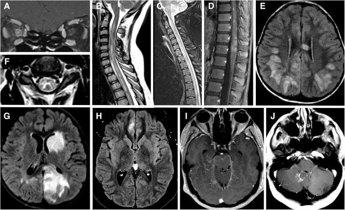
(A) MRI brain T1 coronal post gadolinium contrast showing contrast enhancement of bilateral optic nerves and right optic nerve sheath consistent with perioptic neuritis. (B) MRI spine sagittal STIR showing longitudinal extensive patchy lesion spaning from cervical to thoracic cord. (C) MRI spine sagittal T2 showing hyperintense longitudinally extensive “pseudo-dilation” of central canal. (D) MRI spine sagittal T1 post gadolinium contrast showing patchy enhancement of the conus medullaris. (E) MRI brain axial FLAIR showing large subcortical and septal white matter lesions in a pediatric patient presenting with ADEM. (F) MRI brain axial T2 with hyperintense “H” sign outlining the central gray matter of the upper cervical cord in a teenager with myelitis. (G) MRI brain axial T2 with “fluffy” hyperintense lesion of gray and white matter of the left caudate and left occipital parietal regions in a pediatric patient who presenting with ADEM. (H) MRI brain axial T2 showing unilateral FLAIR hyperintensity and edema of right mesial frontal cortex in a patient with FLAMES syndrome. (I) MRI brain axial T1 post gadolinium contrast showing leptomeningeal enhancement of the midrain and right mesial temporal lobe. (J) MRI brain axial T1 post gadolinium contrast showing a lesion adjacent to the cerebellar vermis and dorsal medulla in a patient with brainstem syndrome and no other lesions.
References
-
- Lebar R, Baudrimont M, Vincent C. Chronic experimental autoimmune encephalomyelitis in the guinea pig. presence of anti-M2 antibodies in central nervous system tissue and the possible role of M2 autoantigen in the induction of the disease. J Autoimmunity. (1989) 2:115–32. 10.1016/0896-8411(89)90149-2 - DOI - PubMed
LinkOut - more resources
Full Text Sources