Review
. 2020 Sep 10:11:656.
doi: 10.3389/fendo.2020.00656. eCollection 2020.

Molecular Mechanisms of Pituitary Cell Plasticity

Affiliations
Review

Molecular Mechanisms of Pituitary Cell Plasticity

Gwen V Childs et al. Front Endocrinol (Lausanne). .

Abstract

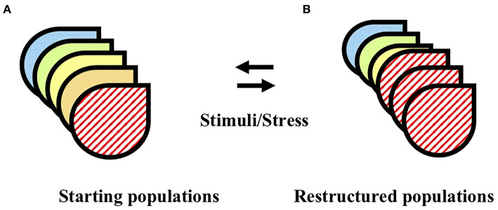
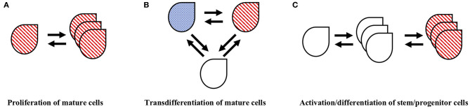
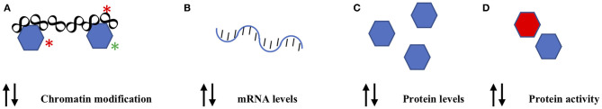
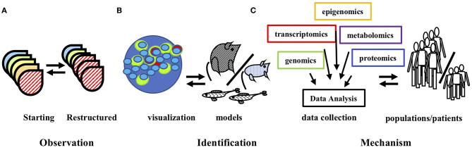
The mechanisms that mediate plasticity in pituitary function have long been a subject of vigorous investigation. Early studies overcame technical barriers and challenged conceptual barriers to identify multipotential and multihormonal cell populations that contribute to diverse pituitary stress responses. Decades of intensive study have challenged the standard model of dedicated, cell type-specific hormone production and have revealed the malleable cellular fates that mediate pituitary responses. Ongoing studies at all levels, from animal physiology to molecular analyses, are identifying the mechanisms underlying this cellular plasticity. This review describes the findings from these studies that utilized state-of-the-art tools and techniques to identify mechanisms of plasticity throughout the pituitary and focuses on the insights brought to our understanding of pituitary function.

Keywords: Musashi; leptin; mRNA translation; multihormonal cells; multipotential; pituitary; plasticity; single cell.


Figures

Figure 1
Figure 1
Pituitary cell populations are restructured in response to physiological stimuli and pathological stressors. (A) The pituitary is comprised of multiple distinct functional cell types (designated by different image fill patterns). (B) In response to physiological stimuli or pathological stress, the pituitary cell populations undergo restructuring. The double arrows imply the potential reversibility of this process. See text for details and relevant references.
Figure 2
Figure 2
Cellular mechanisms that may contribute to pituitary population restructuring. There is evidence that pituitary populations undergo restructuring through multiple cellular mechanisms including (A) mitotic proliferation of existing hormone cells, (B) transdifferentiation of mature hormone cells and (C) activation and differentiation of stem/progenitor cells. The double arrows imply a potential reversibility of these cellular processes. See text for details and relevant references.
Figure 3
Figure 3
Molecular mechanisms that may contribute to pituitary population restructuring. Pituitary cell populations may utilize multiple molecular mechanisms for restructuring including (A) chromatin remodeling (B) gene transcriptional control (C) control of mRNA translation to protein and (D) control of protein function. The double arrows indicate the potential for both activating (positive) and inhibitory (negative) effects upon these molecular mechanisms. See text for details and relevant references.
Figure 4
Figure 4
Technological innovations contribute to understanding pituitary plasticity. (A) Early observations identified remodeling of pituitary hormone cell populations in response to stimuli and stress. (B) Microscopic observations and genetic models identified the cell populations that mediate restructuring. (C) The complimentary use of diverse data set analyses provides insight into the molecular mechanisms of pituitary population restructuring and suggest targets for therapeutic options for patients. The double arrows imply the information feedback of these complimentary technologies. See text for details and relevant references.

References

    1. Le Tissier P, Campos P, Lafont C, Romano N, Hodson DJ, Mollard P. An updated view of hypothalamic-vascular-pituitary unit function and plasticity. Nat Rev Endocrinol. (2017) 13:257–67. 10.1038/nrendo.2016.193 - DOI - PubMed
    1. Fontaine R, Ciani E, Haug TM, Hodne K, Ager-Wick E, Baker DM, et al. Gonadotrope plasticity at cellular, population and structural levels: a comparison between fishes and mammals. Gen Comp Endocrinol. (2020) 287:113344. 10.1016/j.ygcen.2019.113344 - DOI - PubMed
    1. Zhu X, Gleiberman AS, Rosenfeld MG. Molecular physiology of pituitary development: signaling and transcriptional networks. Physiol Rev. (2007) 87:933–63. 10.1152/physrev.00006.2006 - DOI - PubMed
    1. Childs GV, Lloyd JM, Unabia G, Gharib SD, Wierman ME, Chin WW. Detection of luteinizing hormone beta messenger ribonucleic acid (RNA) in individual gonadotropes after castration: use of a new in situ hybridization method with a photobiotinylated complementary RNA probe. Mol Endocrinol. (1987) 1:926–32. 10.1210/mend-1-12-926 - DOI - PubMed
    1. Nunez L, Villalobos C, Senovilla L, Garcia-Sancho J. Multifunctional cells of mouse anterior pituitary reveal a striking sexual dimorphism. J Physiol. (2003) 549(Pt 3):835–43. 10.1113/jphysiol.2003.040758 - DOI - PMC - PubMed

Publication types

LinkOut - more resources