. 2020 Sep 4:11:2020.
doi: 10.3389/fmicb.2020.02020. eCollection 2020.

Evaluation of Neutralizing Antibodies Against Highly Pathogenic Coronaviruses: A Detailed Protocol for a Rapid Evaluation of Neutralizing Antibodies Using Vesicular Stomatitis Virus Pseudovirus-Based Assay

Affiliations

Evaluation of Neutralizing Antibodies Against Highly Pathogenic Coronaviruses: A Detailed Protocol for a Rapid Evaluation of Neutralizing Antibodies Using Vesicular Stomatitis Virus Pseudovirus-Based Assay

Sarah A Almahboub et al. Front Microbiol. .

Abstract

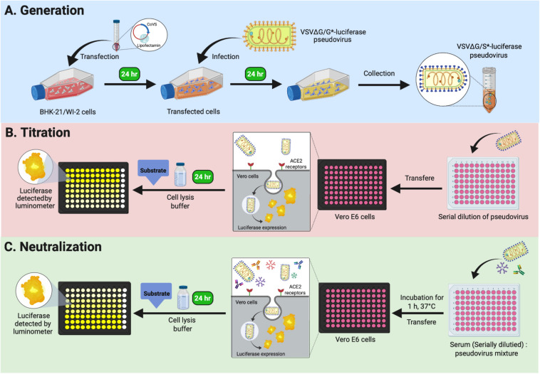
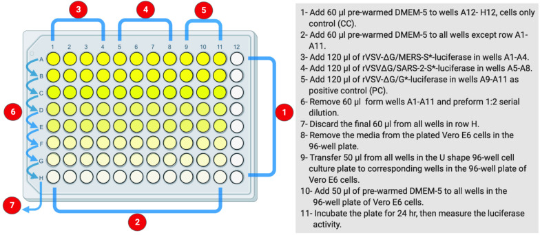
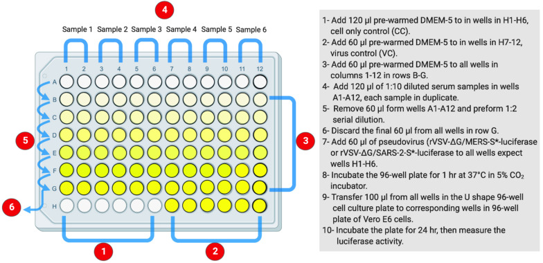
Emerging highly pathogenic human coronaviruses (CoVs) represent a serious ongoing threat to the public health worldwide. The spike (S) proteins of CoVs are surface glycoproteins that facilitate viral entry into host cells via attachment to their respective cellular receptors. The S protein is believed to be a major immunogenic component of CoVs and a target for neutralizing antibodies (nAbs) and most candidate vaccines. Development of a safe and convenient assay is thus urgently needed to determine the prevalence of CoVs nAbs in the population, to study immune response in infected individuals, and to aid in vaccines and viral entry inhibitor evaluation. While live virus-based neutralization assays are used as gold standard serological methods to detect and measure nAbs, handling of highly pathogenic live CoVs requires strict bio-containment conditions in biosafety level-3 (BSL-3) laboratories. On the other hand, use of replication-incompetent pseudoviruses bearing CoVs S proteins could represent a safe and useful method to detect nAbs in serum samples under biosafety level-2 (BSL-2) conditions. Here, we describe a detailed protocol of a safe and convenient assay to generate vesicular stomatitis virus (VSV)-based pseudoviruses to evaluate and measure nAbs against highly pathogenic CoVs. The protocol covers methods to produce VSV pseudovirus bearing the S protein of the Middle East respiratory syndrome-CoV (MERS-CoV) and the severe acute respiratory syndrome-CoV-2 (SARS-CoV-2), pseudovirus titration, and pseudovirus neutralization assay. Such assay could be adapted by different laboratories and researchers working on highly pathogenic CoVs without the need to handle live viruses in the BSL-3 environment.

Keywords: Middle East respiratory syndrome coronavirus; coronaviruses; serological assay; severe acute respiratory syndrome coronavirus 2; vesicular stomatitis virus pseudovirus.


Figures

FIGURE 1
FIGURE 1
Graphical overview of Protocols 1, 2, and 3. (A) Protocol 1: generation of VSV pseudoviruses bearing CoV S protein. (B) Protocol 2: titration assay of the generated VSV pseudoviruses. (C) Protocol 3: neutralization assay to determine CoV-specific nAb titers in serum samples. VSV, vesicular stomatitis virus; CoV, coronavirus; nAb, neutralizing antibody.
FIGURE 2
FIGURE 2
The layout of U-shaped 96-well cell culture plat for rVSV-ΔG/S*-luciferase pseudovirus titration in Protocol 2. The preparation steps are indicated in sequential numbers. rVSV, recombinant vesicular stomatitis virus.
FIGURE 3
FIGURE 3
The layout of U-shaped 96-well cell culture plat for rVSV-ΔG/S*-luciferase pseudovirus neutralization assay in Protocol 3. The preparation steps are indicated in sequential numbers. rVSV, recombinant vesicular stomatitis virus.
FIGURE 4
FIGURE 4
Syncytia formation in BHK-21/WI-2 cells 24 h post-transfection. (A) Cell control. (B) Cells transfected with pCAGGS-G. (C) Cells transfected with pcDNA-SARS-2-S. (D) Cells transfected with pcDNA-MERS-S. Arrows highlight the observed syncytia. Images are representative from three independent experiments.
FIGURE 5
FIGURE 5
Cells rounding 24 h after infection with rVSV-ΔG/G*-luciferase pseudovirus. Cytopathic effect (cells rounding) of BHK-21/WI-2 cells transfected with (A) pCAGGS-G, (B) pcDNA-SARS-2-S, or (C) pcDNA-MERS-S and infected with rVSV-ΔG/G*-luciferase pseudovirus. Images are representative from three independent experiments. rVSV, recombinant vesicular stomatitis virus.
FIGURE 6
FIGURE 6
Neutralization of rVSV pseudovirus generated in the absence or presence of anti-VSV-G polyclonal antibodies. Both rVSV-ΔG/MERS-S*-luciferase and rVSV-ΔG/SARS-2-S*-luciferase were generated in the absence or presence of anti-VSV-G polyclonal antibodies and used in neutralization assay using seropositive serum samples in Vero E6 cells. Using anti-VSV-G polyclonal antibodies always resulted in pseudoviruses without residual rVSV-ΔG/G*-luciferase as shown by the complete inhibition of the luciferase activity compared with the partial inhibition when pseudoviruses were generated in absence of anti-VSV-G polyclonal antibodies, which indicated activities from residual rVSV-ΔG/G*-luciferase. Samples were run in duplicates, and data are shown as mean ± SD from one representative experiment out of three independent experiments. Inhibition (%) was calculated as 100 - [(mean RLU from each sample (virus + diluted serum) - mean RLU from CC)/(mean RLU from VC - mean RLU from CC) × 100]. rVSV, recombinant vesicular stomatitis virus; RLU, relative luminescence unit; CC, cell-only control.
FIGURE 7
FIGURE 7
Titration of rVSV-ΔG/MERS-S*-luciferase and rVSV-ΔG/SARS-2-S*-luciferase pseudoviruses. Generated viruses were serially diluted in a 0.5 log dilution and used to titrate luciferase activity in Vero E6 cells. Luciferase activity was plotted against each dilution using 4PL logistic curve. Samples were run in duplicates, and data are shown as mean ± SD. Data are shown from three different production lots of rVSV-ΔG/MERS-S*-luciferase and rVSV-ΔG/SARS-2-S*-luciferase pseudoviruses. rVSV, recombinant vesicular stomatitis virus.
FIGURE 8
FIGURE 8
Example of neutralization activity using the generated pseudoviruses. Inhibition of rVSV-ΔG/MERS-S*-luciferase, rVSV-ΔG/SARS-2-S*-luciferase, or rVSV-ΔG/G*-luciferase pseudoviruses using seropositive and seronegative human serum samples in Vero E6 cells. Data were plotted using 4PL logistic curves as indicated in section “Materials and Methods.” Serum samples were diluted starting from 1:10 to 1:640. Samples were run in duplicates, and data are shown as mean ± SD from one representative experiment out of three independent experiments. rVSV, recombinant vesicular stomatitis virus.

References

    1. Al-Amri S. S., Abbas A. T., Siddiq L. A., Alghamdi A., Sanki M. A., Al-Muhanna M. K., et al. (2017). Immunogenicity of candidate MERS-CoV DNA vaccines based on the spike protein. Sci. Rep. 7:44875. 10.1038/srep44875 - DOI - PMC - PubMed
    1. Algaissi A., Hashem A. M. (2020). Evaluation of MERS-CoV neutralizing antibodies in sera using live virus microneutralization assay. Methods Mol. Biol. 2099 107–116. 10.1007/978-1-0716-0211-9_9 - DOI - PMC - PubMed
    1. Almasaud A., Alharbi N. K., Hashem A. M. (2020). Generation of MERS-CoV pseudotyped viral particles for the evaluation of neutralizing antibodies in mammalian sera. Methods Mol. Biol. 2099 117–126. 10.1007/978-1-0716-0211-9_10 - DOI - PMC - PubMed
    1. Case J. B., Rothlauf P. W., Chen R. E., Liu Z., Zhao H., Kim A. S. (2020). Neutralizing antibody and soluble ACE2 inhibition of a replication-competent VSV-SARS-CoV-2 and a clinical isolate of SARS-CoV-2. bioRxiv [Preprint]. 10.1101/2020.05.18.102038 - DOI - PMC - PubMed
    1. Cui J., Li F., Shi Z. L. (2019). Origin and evolution of pathogenic coronaviruses. Nat. Rev. Microbiol. 17 181–192. 10.1038/s41579-018-0118-9 - DOI - PMC - PubMed