Review
. 2020 Aug 18:11:1845.
doi: 10.3389/fimmu.2020.01845. eCollection 2020.

Biased MAIT TCR Usage Poised for Limited Antigen Diversity?

Affiliations
Review

Biased MAIT TCR Usage Poised for Limited Antigen Diversity?

Michael N T Souter et al. Front Immunol. .

Abstract

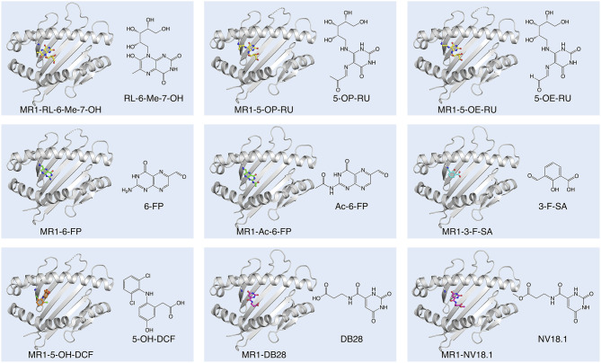
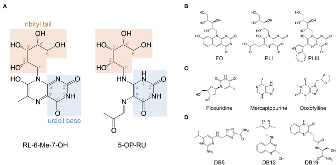
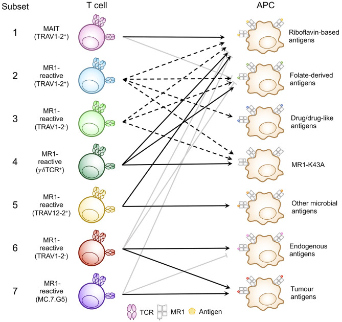
Mucosal-associated invariant T (MAIT) cells are a subset of unconventional T cells that recognize the evolutionarily conserved major histocompatibility complex (MHC) class I-like antigen-presenting molecule known as MHC class I related protein 1 (MR1). Since their rise from obscurity in the early 1990s, the study of MAIT cells has grown substantially, accelerating our fundamental understanding of these cells and their possible roles in immunity. In the context of recent advances, we review here the relationship between MR1, antigen, and TCR usage among MAIT and other MR1-reactive T cells and provide a speculative discussion.

Keywords: MAIT cells; MR1; T cell subsets; TCR repertoire diversity; antigen diversity.


Figures

Figure 1
Figure 1
Diversity of small molecule ligands presented by MR1. Cartoon display (light gray) of the MR1 antigen-binding cleft (top-view) and ball-and-stick display of the antigen (colored) based on the protein data bank (PDB) deposited crystal structures, featuring the human A-F7 MAIT TCR in complex with human MR1-RL-6-Me-7-OH [PDB ID: 4L4V (27)], MR1-5-OP-RU and MR1-5-OE-RU [PDB IDs: 4NQC, 4NQE (26)], MR1-6-FP [PDB ID: 4L4T (27)], MR1-Ac-6-FP [PDB ID: 4PJF (28)], MR1-3-F-SA and MR1-5-OH-DCF [PDB IDs: 5U6Q, 5U72 (29)], and MR1-DB28 and MR1-NV18.1 [PDB IDs:6PVC and 6PVD (30)].
Figure 2
Figure 2
Structures of the riboflavin-based and related MR1 ligands. (A) Chemical structure of 7-hydroxy-6-methyl-8-D-ribityllumazine (RL-6-Me-7-OH) and 5-(2-oxopropylideneamino)-6-D-ribitylaminouracil (5-OP-RU) annotated with the position numbers for relevant functional groups. (B) MR1 ligands isolated from soluble recombinant MR1 expressed with E. coli or M. smegmatis; lumazines 6-(2-carboxyethyl)-7-hydroxy-8-ribityllumazine (photolumazine I; PLI) and 6-(1H-indol-3-yl)-7-hydroxy-8-ribityllumazine (photolumazine III; PLIII) and the riboflavin analog 7,8-didemethyl-8-hydroxy-5-deazariboflavin (FO). (C,D). A selection of MAIT cell agonist and non-agonist MR1 ligands identified from in silico screens of multiple chemical libraries; mercaptopurine, floxuridine, doxofylline, and 2-amino-4-(((3-carbamoyl-1,2,4-oxadiazol-5-l)methyl)amino)-6-isopropylpyrimidin-1-ium (DB5), (4-(((3,5-dimethylisoxazol-4-yl)methyl)amino)quinazolin-2-yl)methanol (DB12) and (S)-N-(1-amino-3-hydroxy-1-oxopropan-2-yl)-3-(3-oxo-3,4-dihydroquinoxalin-2-yl)propenamide (DB19).
Figure 3
Figure 3
Classification of known MR1-reactive T cells based on MR1-antigen reactivity pattern. MR1-reactive T cells bear an αβ or γδ T cell receptor (TCR) that recognizes one or more classes of antigen presented by MR1 on antigen presenting cells (APCs). Solid point arrows indicate that all T cells within the subset recognize the specified antigen, broken point arrows indicate that some T cells within the subset recognize the specified antigen and gray block arrows indicate no recognition of the class of antigen. Table 1 outlines the frequency, TCR usage (where known) and surface characteristics of each subset using this classification. This schematic was inspired by Figure 1 from a recent commentary piece (91).

Similar articles

Cited by

References

    1. Jia Q, Zhou J, Chen G, Shi Y, Yu H, Guan P, et al. . Diversity index of mucosal resident T lymphocyte repertoire predicts clinical prognosis in gastric cancer. Oncoimmunology. (2015) 4:e1001230. 10.1080/2162402X.2014.1001230 - DOI - PMC - PubMed
    1. Shcherbinin DS, Belousov VA, Shugay M. Comprehensive analysis of structural and sequencing data reveals almost unconstrained chain pairing in TCRαβ complex. PLoS Comput Biol. (2020) 16:e1007714. 10.1371/journal.pcbi.1007714 - DOI - PMC - PubMed
    1. Porcelli S, Yockey CE, Brenner MB, Balk SP. Analysis of T cell antigen receptor (TCR) expression by human peripheral blood CD4-8- alpha/beta T cells demonstrates preferential use of several V beta genes and an invariant TCR alpha chain. J Exp Med. (1993) 178:1–16. 10.1084/jem.178.1.1 - DOI - PMC - PubMed
    1. Hashimoto K, Hirai M, Kurosawa Y. A gene outside the human MHC related to classical HLA class I genes. Science. (1995) 269:693–5. 10.1126/science.7624800 - DOI - PubMed
    1. Tilloy F, Treiner E, Park SH, Garcia C, Lemonnier F, De La Salle H, et al. . An invariant T cell receptor alpha chain defines a novel TAP-independent major histocompatibility complex class Ib-restricted alpha/beta T cell subpopulation in mammals. J Exp Med. (1999) 189:1907–21. 10.1084/jem.189.12.1907 - DOI - PMC - PubMed

Publication types

MeSH terms

Substances