. 2020 Sep 9:11:586140.
doi: 10.3389/fpls.2020.586140. eCollection 2020.

Red Light Controls Adventitious Root Regeneration by Modulating Hormone Homeostasis in Picea abies Seedlings

Affiliations

Red Light Controls Adventitious Root Regeneration by Modulating Hormone Homeostasis in Picea abies Seedlings

Sanaria Alallaq et al. Front Plant Sci. .

Abstract

Vegetative propagation relies on the capacity of plants to regenerate de novo adventitious roots (ARs), a quantitative trait controlled by the interaction of endogenous factors, such as hormones and environmental cues among which light plays a central role. However, the physiological and molecular components mediating light cues during AR initiation (ARI) remain largely elusive. Here, we explored the role of red light (RL) on ARI in de-rooted Norway spruce seedlings. We combined investigation of hormone metabolism and gene expression analysis to identify potential signaling pathways. We also performed extensive anatomical characterization to investigate ARI at the cellular level. We showed that in contrast to white light, red light promoted ARI likely by reducing jasmonate (JA) and JA-isoleucine biosynthesis and repressing the accumulation of isopentyl-adenine-type cytokinins. We demonstrated that exogenously applied JA and/or CK inhibit ARI in a dose-dependent manner and found that they possibly act in the same pathway. The negative effect of JA on ARI was confirmed at the histological level. We showed that JA represses the early events of ARI. In conclusion, RL promotes ARI by repressing the accumulation of the wound-induced phytohormones JA and CK.

Keywords: Picea abies; adventitious roots; auxin; conifers; cytokinins; jasmonate; red light; root development.


Figures

Figure 1
Figure 1
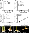
Red light promotes ARI in de-rooted Norway spruce hypocotyls. (A, C) Three-week-old Norway spruce seedlings were de-rooted and kept under cWL for 30 days in in hormone free (HF) distilled water, or in the presence of 1 or 5 μM IAA (A) or in the presence of 1, 5 or 10 μM IBA (B) or NAA (C). (A) Exogenously applied IAA did not have any effect. (B) Three concentrations of IBA did not have any significant effect compared to HF conditions (Repeated measures ANOVA; p = 0.23). (C) Three concentrations of NAA did not have any significant effect compared to HF conditions (Repeated measures ANOVA; p = 0.11). (D) Three-week-old Norway spruce seedlings were de-rooted and kept for 30 days in hormone free (HF) distilled water under cWL or cRL. The hypocotyl cuttings were observed every day to count emerging AR primordia and emerged ARs as in (E). Fifteen seedlings were analyzed for each condition. Adventitious root number increased significantly (t-test, df = 3, p < 0.0001) by cRL compared to cWL over time. (E) Different stages of AR development in de-rooted 3-week-old Norway spruce seedlings in distilled water, under cRL. DAC: days after cut; scale bar = 2 mm.
Figure 2
Figure 2
Light spectral quality controls the homeostasis of endogenous contents of different hormones, which positively or negatively affect ARI. (A–G) Three-week-old Norway spruce seedlings were grown under long day conditions as described in Materials and Methods. De-rooted seedlings were then kept in constant cWL or cRL for 6, 24, 48, and 72 h. For each time point in each condition 5 mm hypocotyls were taken and pooled for hormone quantification. Five independent biological replicates were collected for analysis of IAA, and JA contents and additional five independent biological replicates were collected for CKs quantification. (A) free IAA content; (B) free cis-12-oxo-phytodienoic acid (cis-OPDA) content; (C) free jasmonic acid (JA) content; (D) jasmonate-isoleucine (JA-Ile) content; (E) iP riboside 5′-monophosphate (iPRMP) content; (F) iP riboside content (iPR); (G) isopentyl-adenine (iP) content. Values are the means with standard deviation (SD) of 5 biological replicates. Asterisks indicate statistically significant differences under light conditions cRL versus cWL in t-test; *, **, and *** correspond to P-values of (0.05 > p > 0.01, 0.01 > p > 0.001, and p < 0.001 respectively; n = 5); Dash (#) ndicates statistically significant difference at 24 h, 48 h or 72 h versus 6 h in t-test; p < 0.001, n = 5; FW, fresh weight.
Figure 3
Figure 3
Exogenous application of auxin enhances RL-induced adventitious rooting, whereas exogenously applied CKs and JA repress this process. Three-week-old Norway spruce seedlings were de-rooted and kept under cRL for 30 days in hormone free (HF) distilled water, or in the presence of 1 or 5 μM IBA (A); or in the presence of 1 or 5 µM NAA (B) or in the presence of jasmonate (JA) (C); or in the presence of benzyl adenine (BAP) (D); or in the presence of both JA and IBA (E); or in the presence of both BAP and IBA (F); or in the presence of both JA and BAP (G). (A) All three concentrations of exogenously applied IBA significantly increased the average number of ARs over time (repeated measures ANOVA; P < 0.0001). (B) One or 5 µM of exogenously applied NAA significantly increased the average number of adventitious roots over time (repeated measures ANOVA; p < 0.0001) but no significant improvement compared to HF conditions was observed when 10 μM of NAA were applied (Repeated measures ANOVA; p = 0.48). (C) Exogenously applied JA had a significant negative effect on the average number of ARs over time (repeated measures ANOVA; p < 0.0001). This negative effect was concentration dependent (repeated measures ANOVA; p < 0.0001). (D) Exogenously applied BAP significantly reduced the number of ARs compared to HF conditions (repeated measures ANOVA; p < 0.0001). (E) When JA was applied together with IBA, it significantly repressed the positive effect of IBA. The combination 1 μM IBA + 2 μM JA significantly reduced the number of roots compared to those in HF conditions by 24 days after cut (DAC) and 30 DAC (t-test, degree of freedom df = 14; p = 0.02 and 0.001 respectively). The combination 1 μM IBA + 20 μM JA significantly reduced the average AR number compared to HF conditions (t-test, df = 14; p < 0.01). (F) When BAP was applied together with IBA, it repressed the positive effect of IBA. The combination 1 μM IBA + 0.01 μM BAP significantly reduced the average number of ARs compared to the 1 μM IBA treatment. (13, 15, 18, 24, and 30 DAC; t-test, df = 14; p < 0.009, 0.007, 0.003, 0.0001 and 0.02, respectively). The combination 1 μM IBA + 0.1 μM BAP significantly inhibited rooting induced by 1μM IBA at (13,15,18,24 and 30 DAC (t-test, df = 14; p < 0.008,0.007,0.0009, 0.000000009 and 0.0009 respectively); (G) Both JA and BA repressed AR compared to the HF control, but no significant additive or synergistic effect was observed when JA and BAP were applied together. The combination 0.01 μM BAP + 2 μM JA significantly inhibited rooting compared to the HF control at 24 and 30 DAC (t-test, df = 14; p < 0.0007 and 0.003 respectively), but the result was not significantly different from JA or BAP alone; The combination of 0.01 μM BAP + 5μM JA significantly inhibited rooting compared to the HF controls at 24 and 30 DAC (t-test, df = 14; p < 0.00004 and 0.0000003 respectively), but with no significant difference from JA or BAP alone. (AG) Results are expressed as average number of ARs per cutting. Error bars indicate standard error (SE). For each treatment 15 hypocotyls were used, and the experiment was repeated twice.
Figure 4
Figure 4
JA signaling is downregulated in hypocotyl cuttings kept under constant red light (cRL) compared to hypocotyl cuttings kept under constant white light (cWL). (A) Relative transcript amount of putative JA-responsive genes PaMYC2-like, PaJAZ3-like, PaJAZ10-like and PaAOC-like in 3-week-old Norway spruce seedling hypocotyls treated for 3 h with 50 μM JA or with a mock treatment. Values are relative (on a log10 scale) to the values for mock treated seedlings which were arbitrarily set to 1. Error bars indicate ±SE obtained from two independent biological replicates, with three technical replicates of each. A t-test was carried out, and values indicated by asterisks differed significantly from the mock values (p < 0.001, n ≥ 80). (B) Relative transcript amounts of putative JA-responsive genes PaMYC2-like, PaJAZ3-like, PaJAZ10-like and PaAOC-like, and the putative JA receptor PaCOI1-like, at the base of 3-week-old de-rooted hypocotyls kept under cRL or cWL for 6, 24, 48 and 72 h. The values are relative (on a log2 scale) to the values obtained from seedlings kept under cWL, which were arbitrarily set to 1. Error bars indicate ±SE obtained from two independent biological replicates, with three technical replicates of each. A t-test was carried out, and asterisks indicate values that differed significantly from the control values (p < 0.001, n ≥ 80).
Figure 5
Figure 5
Histological analysis of AR development in Norway spruce hypocotyl cuttings under cRL. (A) Cross section through the base of a 21-day-old hypocotyl cutting just before transfer to rooting conditions. (B) Longitudinal section through the base of a hypocotyl cutting kept in hormone free (HF) conditions for 5 days. Arrowheads indicate localized cell divisions, probably at the points of origin of future ARs. (C) Longitudinal section through the base of a hypocotyl cutting kept in the presence of 1 μM IBA for 10 days. Arrowheads indicate the presence of clusters of dividing cells. (D) Longitudinal section through the base of a hypocotyl cutting kept in the presence of 1 μM IBA for 13 days. Arrows indicate an adventitious root primordium (ARP) and an emerged adventitious root (AR). (E) Cross section through the base of a hypocotyl cutting kept in HF conditions for 15 days. Arrows indicate an ARP and an emerged AR. e, epidermis; Co, cortex; Pa, parenchyma; Ph, phloem; en, endodermis; c, cambium region; P, pith; x, xylem. In all panels scale bars = 20 μm.
Figure 6
Figure 6
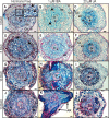
Histological analysis of ARI in Norway spruce hypocotyl cuttings under cRL in the presence of IBA, JA or HF. (A, D, G, J) Cross sections were prepared from the base of de-rooted Norway spruce hypocotyl cuttings, kept under cRL in hormone free (HF) distilled water for 3, 5, 10, or 13 days. In (A), the arrows show early cell activation, i.e. small cells with dense cytoplasm and large nuclei. In (D), arrows indicate anticlinal and periclinal division. In (G) arrows show the presence of tracheal elements. In (J), arrows indicate clusters of dividing cells developing at the periphery of the tracheal elements. (B, E, H, K) Cross sections were taken from the base of de-rooted Norway spruce hypocotyl cuttings, kept under cRL in the presence of 1 μM IBA for 3, 5, 10, or 13 days. In (B) arrows indicate cell divisions; In (H) arrows indicate the presence of clusters of dividing cells for which close-up views are shown. In (K) arrows indicate emerged adventitious roots (ARs). (C, F, I, L) Cross sections were prepared from the base of de-rooted Norway spruce hypocotyl cuttings, kept under cRL in the presence of 20 μM JA for 3, 5, 10, or 13 days. e, epidermis; Co, cortex; Ph, phloem; en, endodermis; c, cambium region; P, pith; x, xylem. In all panels scale bars = 20 μm.

Similar articles

Cited by

References

    1. Abarca D., Díaz-Sala C. (2009). “Adventitious root formation in conifers,” in Adventitious Root Formation of Forest Trees and Horticultural Plants – from Genes to Applications. Eds. Niemi K., Scagel C. (Kerala, India: Research Signpost Publishers; ).
    1. Agulló-Antón M.Á., Sánchez-Bravo J., Acosta M., Druege U. (2011). Auxins or Sugars: What Makes the Difference in the Adventitious Rooting of Stored Carnation Cuttings? J. Plant Growth Regul. 30, 100–113. 10.1007/s00344-010-9174-8 - DOI
    1. Alallaq S., Ranjan A., Brunoni F., Novák O., Lakehal A., Bellini C. (2020). Red light controls adventitious root regeneration by modulating hormone homeostasis in Picea abies seedlings. BioRxiv. 10.1101/2020.03.11.985838 - DOI - PMC - PubMed
    1. Antoniadi I., Plačková L., Simonovik B., Doležal K., Turnbull C., Ljung K., et al. (2015). Cell-type-specific cytokinin distribution within the arabidopsis primary root apex. Plant Cell 27, 1955–1967. 10.1105/tpc.15.00176 - DOI - PMC - PubMed
    1. Baque M. A., Hahn E.-J., Paek K.-Y. (2010). Induction mechanism of adventitious root from leaf explants of Morinda citrifolia as affected by auxin and light quality. Vitr. Cell. Dev. Biol. - Plant 46, 71–80. 10.1007/s11627-009-9261-3 - DOI