. 2020 Sep 18:2020:7416493.
doi: 10.1155/2020/7416493. eCollection 2020.

Isolation and Characterization of Human Synovial Fluid-Derived Mesenchymal Stromal Cells from Popliteal Cyst

Affiliations

Isolation and Characterization of Human Synovial Fluid-Derived Mesenchymal Stromal Cells from Popliteal Cyst

Fang Li et al. Stem Cells Int. .

Abstract

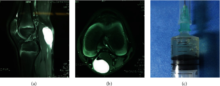
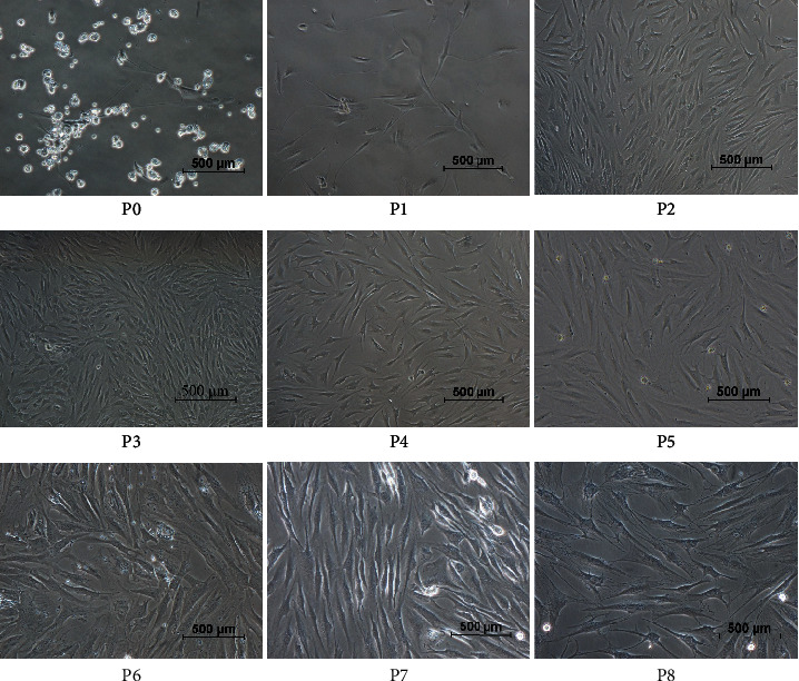
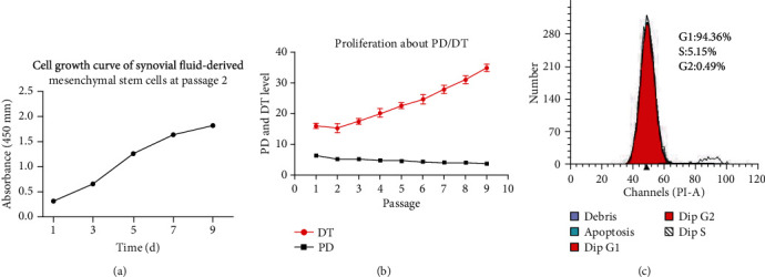
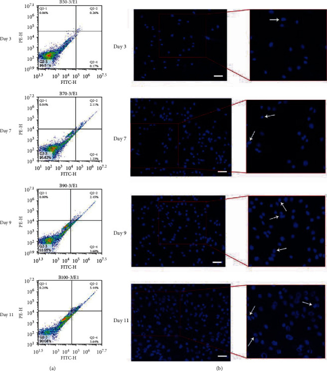
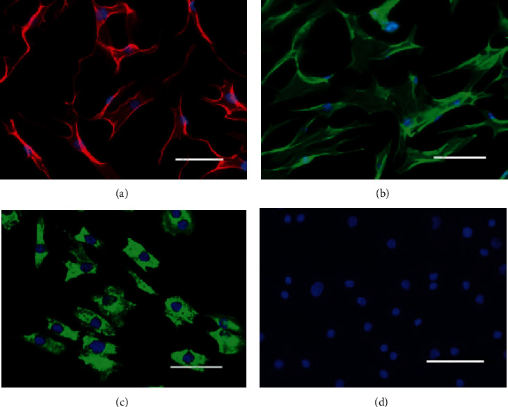
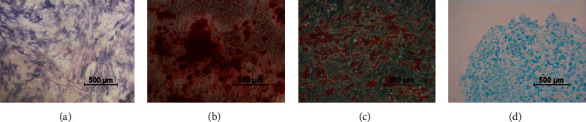
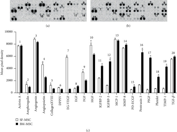
Mesenchymal stem cells (MSCs) are multipotent progenitor cells in adult tissues. The aim of this study is to isolate and identify synovial fluid-derived mesenchymal stromal cells (SF-MSCs) from the popliteal cyst fluid of pediatric patients. SF-MSCs were collected from the popliteal cyst fluid of pediatric patients during cystectomy surgery. After cyst fluid extraction and adherent culturing, in vitro morphology, growth curve, and cell cycle were observed. The expression of stem cell surface markers was analyzed by flow cytometry, and expression of cell marker protein was detected by immunofluorescence. SF-MSCs were cultured in osteogenic, adipogenic, and chondrogenic differentiation medium. The differentiation potential of SF-MSCs was analyzed by alkaline phosphatase (Alizarin Red), Oil Red O, and Alcian blue. Antibody detection of human angiogenesis-related proteins was performed compared with bone marrow mesenchymal stem cells (BM-MSCs). The results show that SF-MSCs from the popliteal cyst fluid of pediatric patients showed a shuttle appearance and logarithmic growth. Flow cytometry analysis revealed that SF-MSCs were negative for hematopoietic lineage markers (CD34, CD45) and positive for MSC markers (CD44, CD73, CD90, and CD105). Interstitial cell marker (vimentin) and myofibroblast-like cell marker alpha-smooth muscle actin (α-SMA) were positive. These cells could differentiate into osteogenic, adipogenic, and chondrogenic lineages, respectively. Several types of human angiogenesis-related proteins were detected in the cell secretory fluid. These results show that we successfully obtained SF-MSCs from the popliteal cyst fluid of pediatric patients, which have the potential to be a valuable source of MSCs.


Conflict of interest statement

The authors declare no competing interests.

Figures

Figure 1
Figure 1
(a) T2-weighted sagittal scan and (b) T2-weighted axial scan of the right knee showing a cystic lesion between the semimembranous muscle and the medial head of the gastrocnemius. The popliteal cyst is filled with liquid. (c) Extraction fluid of popliteal cyst: it shows a colorless mucous liquid with little blood color.
Figure 2
Figure 2
Morphological characteristics of SF-MSCs. P0: representative characteristics of SF-MSCs at primary passage within 48 h—the cell exhibiting individual colony adhered to the culture plates and nonadherent or few adherent small round cells are scattered in the culture plates (100x). P1-P4: representative characteristics of SF-MSCs from passage 1 to passage 3, between day 3 and day 10—cells exhibit the fibroblastic spindle-like shape (100x). P5-P8: representative characteristics of SF-MSCs from passage 4 to passage 8—the cells show fibrocyte-like form with long fusiform shape, grow in the same direction, and maintain the morphology (100x).
Figure 3
Figure 3
(a) SF-MSC growth curve: cells showed normal exponential cell growth patterns with a steady increase in number during an 11-day culture period. The determination and calculation of the doubling time to assess proliferation (CCK-8) showed that the growth rate was slow during the first 3 days and rapid from 4 to 8 days. The cells reached the platform stage (stopped growing or entered apoptosis) at 9~11 days. These cells showed normal exponential cell growth patterns with a steady increase in number during an 11-day culture period, as well as general mesenchymal stem cell features. (b) Curve diagram of population doublings and doubling time with passages of rUSCs. (c) Cell cycle of SF-MSCs: the data showed that 94.36% of cells were in G0/G1 phase, 0.49% were in G2/M phase, and only 5.15% were in the S phase, indicating that the cells have strong proliferative abilities.
Figure 4
Figure 4
Cell apoptosis analyses of SF-MSCs after culture for 3, 7, 9, and 11 days. (a) Apoptotic cells were quantified by double-supravital staining with Annexin V-FITC and PI. (b) Cells were stained with Hoechst 33342 and were examined under a fluorescence microscope at magnification of ×200 (scale bar = 50 μm). The arrows indicate several apoptotic cells with typical condensation of chromatin, cell shrinkage, and nuclear fragmentation.
Figure 5
Figure 5
Surface marker expression of CD24, CD29, CD34, CD44, CD45, CD73, CD90, CD105, CD117, CD146, CD147, and OCT-4 in SF-MSCs after 3rd passage from the popliteal cyst analyzed by flow cytometry. Note: FITC: fluorescein isothiocyanate; PE: phycoerythrin.
Figure 6
Figure 6
Microscopic image showing the immunofluorescent staining of SF-MSCs at passage 3. (a) Immunofluorescent staining of SF-MSCs showing positive expression of vimentin, (b) immunofluorescent staining of SF-MSCs showing positive expression of alpha-SMA, (c) immunofluorescent staining of SF-MSCs showing positive expression of collagen I, and (d) immunofluorescent staining of SF-MSCs showing negative expression of pan keratin. The nuclei were counterstained with DAPI. Scale bars = 200 μm.
Figure 7
Figure 7
Differentiation characteristics of SF-MSCs at passage 2. (a) Representative of early osteogenesis was detected by alkaline phosphatase staining at day 7. (b) Representative of osteogenesis detected as calcium deposition is an indication of osteogenesis and was detected using Alizarin Red at day 22. (c) Representative of adipogenic differentiation visualized by Oil Red O staining of the intracellular lipid vesicles at day 21. (d) Representative of chondrogenic differentiation detected by Alcian blue staining at day 28. Micrographs are representative of 12 experiments.
Figure 8
Figure 8
Angiogenesis-related protein growth factor secretion of SF-MSCs and BM-MSCs. (a) Result of angiogenesis growth factor secretion of SF-MSCs, (b) result of angiogenesis growth factor secretion of BM-MSCs, and (c) statistical analysis of secretion of SF-MSC and BM MSC by using Student's t-test: P < 0.05, SF-MSC versus BM-MSC.
Figure 9
Figure 9
Histological assessment of cyst wall tissue. The HE staining (a) and Masson staining (b) of cyst wall tissue show the presence of endothelial cells of blood vessels in popliteal cyst. Immunohistochemical staining of CD31 (c) and vWF (d) showed positive expression. Immunohistochemical staining of inside wall tissue of popliteal cyst cavity showed negative expression of AE1/3 (e). Scale bars = 200 μm.

Similar articles

Cited by

References

    1. Alison M. R., Poulsom R., Jeffery R., et al. Hepatocytes from non-hepatic adult stem cells. Nature. 2000;406(6793):p. 257. doi: 10.1038/35018642. - DOI - PubMed
    1. Jones B. A., Pei M. Synovium-Derived Stem Cells: A Tissue-Specific Stem Cell for Cartilage Engineering and Regeneration. Tissue Engineering. Part B, Reviews. 2012;18(4):301–311. doi: 10.1089/ten.teb.2012.0002. - DOI - PubMed
    1. Wakitani S., Mitsuoka T., Nakamura N., Toritsuka Y., Nakamura Y., Horibe S. Autologous bone marrow Stromal cell transplantation for repair of full-thickness articular cartilage defects in Human Patellae: two case reports. Cell Transplantation. 2017;13(5):595–600. doi: 10.3727/000000004783983747. - DOI - PubMed
    1. Toh W. S., Lai R. C., Hui J. H. P., Lim S. K. MSC exosome as a cell-free MSC therapy for cartilage regeneration: implications for osteoarthritis treatment. Seminars in Cell & Developmental Biology. 2017;67:56–64. doi: 10.1016/j.semcdb.2016.11.008. - DOI - PubMed
    1. Kuroda R., Usas A., Kubo S., et al. Cartilage repair using bone morphogenetic protein 4 and muscle-derived stem cells. Arthritis and Rheumatism. 2006;54(2):433–442. doi: 10.1002/art.21632. - DOI - PubMed