. 2020 Sep 18:2020:8883436.
doi: 10.1155/2020/8883436. eCollection 2020.

p130 And pRb in the Maintenance of Transient Quiescence of Mesenchymal Stem Cells

Affiliations

p130 And pRb in the Maintenance of Transient Quiescence of Mesenchymal Stem Cells

Boris Popov et al. Stem Cells Int. .

Abstract

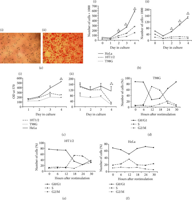
An effective regulation of quiescence plays a key role in the differentiation, plasticity, and prevention of stem cells from becoming malignant. The state of quiescence is being controlled by the pRb family proteins which show overlapping functions in cell cycle regulation; however, their roles in controlling the proliferation of mesenchymal stem cells (MSCs) remain to be understood. This study investigated the regulation of transient quiescence using growth curves, proliferation assay, the cytometric evaluation of cell cycle, Western blotting, and the electromobility gel shift assay (EMSA) on synchronized MSCs of the C3H10Т1/2 and control cells with different statuses of pRb proteins. It has been found that functional steady-state level of p130 but not pRb plays a critical role for entering, exiting, and maintenance of transient quiescence in multipotent mesenchymal stem cells.


Conflict of interest statement

No conflicts of interest, financial, or otherwise are declared by all authors.

Figures

Figure 1
Figure 1
MSCs of the 10T1/2 cell line show serum-dependent growth restriction and cell cycle-dependent oscillation. (a) 10T1/2 cell line possesses the ability to adipocyte differentiation. The cells were observed before AD (i) and in 10 days (ii) after the induction of AD and Red Oil staining under the Pascal microscope (Carl Zeiss, Germany) with transmitted light using a 10x objective. (b) Growth curves of T98G, 10T1/2, and HeLa cell lines: i—full growth medium, ii—serum-deprived medium. Equal cell numbers of each line were plated into wells of 96 cell culture plate in triplicate and counted every day until day 4. The data are presented as mean ± SD, p < 0.01 compared to the 10T1/2 cells, ∗∗p < 0.01 compared to the T98G cells. (c) Cell proliferation rate of the 10T1/2 cells: i—full growth medium, ii—serum-deprived medium. The cell proliferation rate was estimated using the MTT assay. The asynchronously growing 10T1/2, T98G, and HeLa cells were seeded at a concentration of 2 × 10⁴ cells/well in 100 μl full culture medium in 24 wells of 96-well cell culture plate and incubated for 24 h. Then, 12 wells containing cells of each cell line were filled with full growth medium (FM) while the other 12—with serum-free medium (SFM). For the next four days, the cells were treated with the MTT reagent followed by measuring OD by ELISA reader at 570 nm. The data are presented as mean ± SD, p < 0.05 compared to the 10T1/2 cells, ∗∗p < 0.05 compared to the T98G cells. (d–f) Flow cytometry analysis of cell cycle progression of T98G, MSCs, and HeLa cell lines, accordingly. The cells were cultured for 72 h in DMEM with 0.15% FCS, restimulated with 10% FCS, and used for flow cytometry in 6 h intervals after restimulation.
Figure 2
Figure 2
Western blotting analysis of the p130 and pRb cell cycle-dependent oscillations in 10T1/2, T98G, and HeLa cell lines. The cells of all cell lines were synchronized as described above. The cell extracts containing 40 μg of total protein were resolved on 8% SDS–PAGE for the following Western blotting: (a) T98G cell line; (b) MSCs (10 T1/2 cell line); (c) HeLa cell line. The data are presented as mean ± SD.
Figure 3
Figure 3
The analysis of the protein structure of the pocket proteins (pp)-E2f-DNA complexes in the T98G, HeLa, and the 293HEK cells by EMSA. (a) The proteins composing the pp-E2f-DNA complexes in the quiescent and cycling T98G cells. (b) The structure of the pp-E2f-DNA complexes in the cycling HeLa cells. (c) The pp-E2f-DNA complexes are completely absent in the 293HEK cells. The samples prepared from the cycling cells were treated with competitor—C, deoxycholate—D, or different antibodies as indicated. The cycling T98G, HeLa, and 293HEK cells were used, accordingly, in the 18 h and 12 h points after restimulation. The protein-DNA bands that included different proteins designated with (+), the band abolishment—(-), the supershifts induced by specific antibodies—(++), and nonspecific bands—(∗).
Figure 4
Figure 4
The structure of the pp-E2f-DNA complexes in quiescent 10T1/2 cells. (a) The pp-E2f4-DNA complexes in the quiescent and cycling T98G, 10T1/2, and HeLa cell lines. (b) Analysis of protein structure of the pp-E2f-DNA complexes in the quiescent 10T1/2 cells. The cells were synchronized as described above. The abbreviations are the same as in Figure 3.

Similar articles

References

    1. Cobrinik D. Pocket proteins and cell cycle control. Oncogene. 2005;24(17):2796–2809. doi: 10.1038/sj.onc.1208619. - DOI - PubMed
    1. Sage J., Mulligan G. J., Attardi L. D., et al. Targeted disruption of the three Rb-related genes leads to loss of G(1) control and immortalization. Genes and Development. 2000;14(23):3037–3050. doi: 10.1101/gad.843200. - DOI - PMC - PubMed
    1. Burkhart D. L., Sage J. Cellular mechanisms of tumour suppression by the retinoblastoma gene. Nature Reviews Cancer. 2008;8(9):671–682. doi: 10.1038/nrc2399. - DOI - PMC - PubMed
    1. Hurford R. K., Jr., Cobrinik D., Lee M. H., Dyson N. pRB and p107/p130 are required for the regulated expression of different sets of E2F responsive genes. Genes and Development. 1997;11(11):1447–1463. doi: 10.1101/gad.11.11.1447. - DOI - PubMed
    1. Ishida S., Huang E., Zuzan H., et al. Role for E2F in control of both DNA replication and mitotic functions as revealed from DNA microarray analysis. Molecular and Cellular Biology. 2001;21(14):4684–4699. doi: 10.1128/MCB.21.14.4684-4699.2001. - DOI - PMC - PubMed

LinkOut - more resources