

Pulmonary artery aneurysm as a result of group 2 pulmonary artery hypertension in a patient with significant mitral and aortic valve disease - review and case report
- PMID: 33014090
- PMCID: PMC7526489
- DOI: 10.5114/kitp.2020.99078
Pulmonary artery aneurysm as a result of group 2 pulmonary artery hypertension in a patient with significant mitral and aortic valve disease - review and case report
Abstract
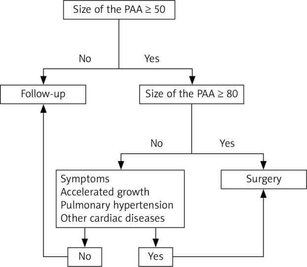
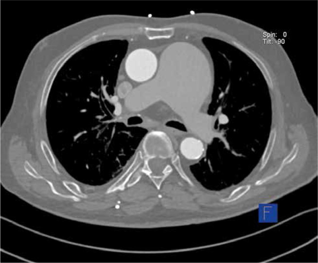
Aneurysms are uncommon, but potentially life-threatening abnormalities of the pulmonary arteries. Aneurysm of the main pulmonary artery (MPA) defined as MPA diameter over 40 mm was reported in 1 : 14 000 autopsies. The most frequent location is the main pulmonary artery (89% of cases), whereas the maximum described diameter is 106-170 mm. Clinical manifestations are usually nonspecific or asymptomatic. Right heart failure symptoms, pulmonary regurgitation, trachea or bronchi compression or pulmonary emboli caused by enlarged MPA are the most commonly described clinical manifestations. Pulmonary artery aneurysm dissection is an uncommon complication but associated with a high mortality rate. Unfortunately, guidelines regulating the optimal time for the surgical intervention still have not been developed. We present the history of 76-year-old patient suffering from an aneurysm of the pulmonary artery (74 × 61 mm), as well as mitral and aortic valve disease, who was successfully operated on in our hospital.
Tętniaki są rzadkimi, ale potencjalnie zagrażającymi życiu patologiami tętnicy płucnej. Częstość ich występowania szacuje się na 1 : 14 000 autopsji. Zgodnie z definicją tętniaka pnia tętnicy płucnej rozpoznaje się, gdy średnica naczynia przekracza 40 mm. Najczęstszą jego lokalizacją jest pień płucny (89% przypadków), podczas gdy największy opisany dotąd tętniak miał średnicę 106–170 mm. Objawy są najczęściej niespecyficzne lub choroba przebiega bezobjawowo. Prawokomorowa niewydolność serca, niedomykalność zastawki płucnej, ucisk na tchawicę, oskrzela lub zator płucny spowodowany przez ucisk powiększającego się tętniaka są najczęstszymi objawami zgłaszanymi przez chorego. Rozwarstwienie tętniaka tętnicy płucnej jest bardzo rzadkim powikłaniem, ale obarczonym wysoką śmiertelnością. Niestety dotychczas nie zostały opracowane żadne wytyczne dotyczące postępowania u pacjentów z tą chorobą. Przedstawiamy przypadek 76-letniego pacjenta z tętniakiem tętnicy płucnej (74 × 61 mm), wadą zastawki aortalnej i mitralnej, który został zoperowany w naszym ośrodku.
Keywords: pulmonary artery aneurysm; pulmonary hypertension.
Copyright © 2020 Polish Society of Cardiothoracic Surgeons (Polskie Towarzystwo KardioTorakochirurgów) and the editors of the Polish Journal of Cardio-Thoracic Surgery (Kardiochirurgia i Torakochirurgia Polska).
Conflict of interest statement
The authors report no conflict of interest.
Figures


References
-
- Deterling RA Jr, Clagett OT. Aneurysm of the pulmonary artery: review literature and report of a case. Am Heart J 1947; 34: 471-499. - PubMed
-
- Akagi S, Nakamura K, Sarashina T, et al. Progression of pulmonary artery dilatation in patients with pulmonary hypertension coexisting with a pulmonary artery aneurysm. J Cardiol 2018; 71: 517-522. - PubMed
-
- Duijnhouwer AL, Navarese EP, Van Dijk APJ, et al. Aneurysm of the pulmonary artery, a systematic review and critical analysis of current literature. Congenit Heart Dis 2016; 11: 102-109. - PubMed
-
- Kanaoka K, Horii M, Nagato H, Kaneda K. A giant pulmonary artery aneurysm. Eur Heart J Cardiovasc Imaging 2018; 19: 236. - PubMed
Publication types
LinkOut - more resources
Full Text Sources