Review
. 2020 Sep;17(3):155-159.
doi: 10.5114/kitp.2020.99080. Epub 2020 Sep 23.

Useful assessment of myocardial viability and dyssynchrony from gated perfusion scintigraphy for better qualification for resynchronization therapy. Part 3

Affiliations
Review

Useful assessment of myocardial viability and dyssynchrony from gated perfusion scintigraphy for better qualification for resynchronization therapy. Part 3

Monika Z Czaja-Ziółkowska et al. Kardiochir Torakochirurgia Pol. 2020 Sep.

Abstract

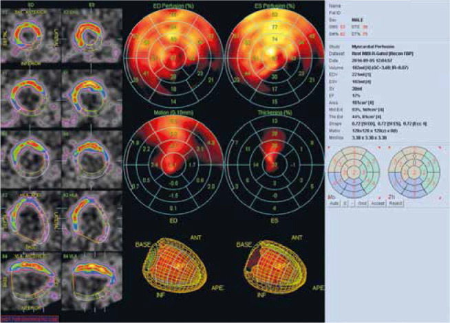
The first part of the review concerning myocardial imaging by single photon emission computed tomography (SPECT) discussed the basic aspects of interpretation of left ventricular perfusion disorders in stress and rest examination. The second part presented the interpretation of gated SPECT imaging in relation to the assessment of systolic and diastolic left ventricular functions. The third part concerns the assessment of myocardial viability and phase analysis from gated SPECT in the qualification of patients with left ventricular systolic dysfunction for cardiac resynchronization therapy.

W pierwszej części cyklu prac dotyczących scyntygrafii perfuzyjnej mięśnia sercowego metodą tomografii emisyjnej pojedyn- czego fotonu (SPECT) omówiono podstawowe zagadnienia na temat interpretacji zaburzeń perfuzji lewej komory w rejestracji wysiłkowej i spoczynkowej. W drugiej części przedstawiono interpretacje badania bramkowanego zapisem elektrokardiograficznym (GATED-SPECT) w odniesieniu do oceny funkcji skurczowej i rozkurczowej lewej komory. Trzecia część została poświęcona ocenie żywotności mięśnia sercowego oraz analizie fazowej badania bramkowanego w kwalifikacji pacjentów z dysfunkcją skurczową lewej komory do terapii resynchronizującej.

Keywords: GATED-SPECT; myocardial viability; phase analysis.


Conflict of interest statement

The authors report no conflict of interest.

Figures

Figure 1
Figure 1
Assessment of left ventricular myocardial viability in patient A. Lack of viability of the inferior and lateral wall of the left ventricle and preserved viability of the anterior wall, septum and apex in the resting myocardial perfusion SPECT examination with attenuation correction. The polar map divided into 17 segments. The examination was performed in the Laboratory of Nuclear Medicine of the Silesian Center for Heart Diseases in Zabrze (Cedars-Sinai Medical Center software; QPS – Quantified Perfusion SPECT)
Figure 2
Figure 2
Lack of perfusion in patient A. The same patient as in Figure 1. The figure shows the extent of perfusion defects in the resting myocardial perfusion SPECT study with attenuation correction. Polar map division based on arteries. The perfusion deficits correspond to the right coronary artery and the circumflex branch. The examination was performed in the Laboratory of Nuclear Medicine of the Silesian Center for Heart Diseases in Zabrze (Cedars-Sinai Medical Center software; QPS – Quantified Perfusion SPECT)
Figure 3
Figure 3
Point assessment of myocardial viability in patient A. The same patient as in Figures 1, 2. Rest myocardial perfusion SPECT examination with attenuation correction. The polar map divided into 17 segments. According to the point scale, all segments of the inferior and lateral wall were assigned a value of myocardial perfusion defects of 2 or 3 points, which means that there are no clear intermediate features of preserved muscle viability in this area. The assessment of muscle viability should also take into account the result of the left ventricular systolic thickening from GATED-SPECT imaging. The examination was performed in the Laboratory of Nuclear Medicine of the Silesian Center for Heart Diseases in Zabrze (Cedars-Sinai Medical Center software; QPS – Quantified Perfusion SPECT)
Figure 4
Figure 4
Contractility abnormalities and impaired thickening in patient A. The same patient as in Figures 1–3. In the GATEDSPECT resting examination, global contractility abnormalities and impaired systolic thickening of the left ventricle were observed. The polar map divided into 17 segments. Akinesis/dyskinesis of the inferior wall and hypokinesis/akinesis of the lateral wall and the lack of systolic thickness in these areas confirm the lack of myocardial viability in the right coronary artery and the circumflex branch territories. Contractility disorders in the anterior wall and septum as well as the apex with the absence of significant perfusion disorders in these areas indicate the presence of “myocardial hibernation” (significant stenosis of the left main coronary artery in coronary angiography). The examination was performed in the Laboratory of Nuclear Medicine of the Silesian Center for Heart Diseases in Zabrze (Cedars-Sinai Medical Center software; QGS – Quantified Gated SPECT)
Figure 5
Figure 5
Assessment of left ventricular mechanical dyssynchrony. Significantly increased end-diastolic and end-systolic volumes of the left ventricle with a significant reduction in LV ejection fraction (about 17%). Phase analysis indicates a high degree of dyssynchrony of the contraction in the infero-lateral wall. PHB is 40°, PSD 13°, and Entropy 72%. The result of the study indicates that the patient is a good candidate for CRT implantation. The study was performed in the Laboratory of Nuclear Medicine of the Silesian Center for Heart Diseases in Zabrze (Cedars-Sinai Medical Center software; QGS – Quantified Gated SPECT) PHB – phase histogram bandwidth, PSD – phase standard deviation.
Figure 6
Figure 6
CRT non-responders phase analysis. An example of a patient without response to CRT. Bottom: segment 6 and 7 phase analysis. PHB, PSD and Entropy for segment 6 were 36°, 10.5° and 45%, respectively, for segment 7: 18°, 4.2° and 26% respectively. Patient with left bundle branch block and left ventricular ejection fraction below 35%. The study was performed in the Laboratory of Nuclear Medicine of the Silesian Center for Heart Diseases in Zabrze (Cedars-Sinai Medical Center software; QGS – Quantified Gated SPECT) PHB – phase histogram bandwidth, PSD – phase standard deviation.
Figure 7
Figure 7
Left ventricular activation map. The figure shows the map of left ventricular activation. In this example, the latest activation was located in the LV lateral wall region (mean phase 183°). The study was performed in the Laboratory of Nuclear Medicine of the Silesian Center for Heart Diseases in Zabrze (Cedars-Sinai Medical Center software; QGS – Quantified Gated SPECT)

Similar articles

Cited by

References

    1. Czaja M, Wygoda Z, Duszańska A, et al. . Interpreting myocardial perfusion scintigraphy using single-photon emission computed tomography. Part 1. Kardiochir Torakochir Pol 2017; 14: 192-199. - PMC - PubMed
    1. Czaja M, Wygoda Z, Duszańska A, et al. . Myocardial perfusion scintigraphy–interpretation of gated imaging. Part 2. Kardiochir Torakochir Pol 2018; 15: 49-56. - PMC - PubMed
    1. Ponikowski P, Voors AA, Anker SD, et al. ; 2016 ESC Guidelines for the diagnosis and treatment of acute and chronic heart failure: The TaskForce for the diagnosis and treatment of acute and chronic heart failure of the European Society of Cardiology (ESC). Developed with the special contribution of the Heart Failure Association (HFA) of the ESC. Eur J Heart Fail 2016; 18: 891-975. - PubMed
    1. Petretta M, Storto G, Acampa W, et al. . Relation between wall thickening on gated perfusion SPECT and functional recovery after coronary revascularization in patients with previous myocardial infarction. Eur J Nucl Med Mol Imaging 2004; 31: 1599-1605. - PubMed
    1. Nakajima K, Matsumoto N, Kasai T, et al. . Normal values and standardization of parameters in nuclear cardiology: Japanese Society of Nuclear Medicine working group database. Ann Nucl Med 2016; 30: 188-199. - PMC - PubMed