

Percutaneous closure of large pulmonary artery to left atrial fistula
- PMID: 33014197
- PMCID: PMC7520539
- DOI: 10.1016/j.jccase.2020.05.017
Percutaneous closure of large pulmonary artery to left atrial fistula
Abstract
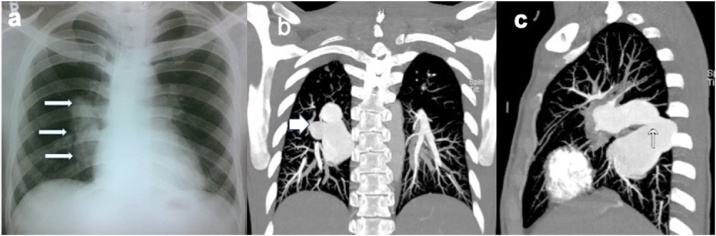
Pulmonary artery to the left atrial fistula is a rare anomaly. Two young males presented with effort intolerance and cyanosis and had large pulmonary artery to left atrial fistula from right and left pulmonary artery, respectively. The right-sided fistula was closed with a large duct occluder device in transseptal approach whereas the left-sided fistula was closed with a large muscular ventricular septal device. Complete occlusion and symptomatic relief was achieved in both cases. In the follow up the patients were doing well.
Keywords: Cyanosis; Device closure; Fistula; Left atrium; Pulmonary artery.
© 2020 Japanese College of Cardiology. Published by Elsevier Ltd. All rights reserved.
Figures


Similar articles
-
Device closure of fistula from left lower pulmonary artery to left atrium using a vascular plug: A case report.World J Cardiol. 2021 Apr 26;13(4):111-116. doi: 10.4330/wjc.v13.i4.111. World J Cardiol. 2021. PMID: 33968310 Free PMC article.
-
A large type I right pulmonary artery to left atrium fistula: underwent successful percutaneous device closure with duct occluder-a rare case report.Egypt Heart J. 2024 Feb 21;76(1):24. doi: 10.1186/s43044-024-00438-w. Egypt Heart J. 2024. PMID: 38381361 Free PMC article.
-
Transcathether closure of a right pulmonary artery-to-left atrial fistula using Amplatzer muscular ventricular septal defect occluder.Cardiovasc Interv Ther. 2014 Oct;29(4):359-62. doi: 10.1007/s12928-013-0240-6. Epub 2014 Jan 4. Cardiovasc Interv Ther. 2014. PMID: 24390925
-
Surgical treatment of a fistula between the right pulmonary artery and the left atrium: presentation of two cases and review of literature.Eur J Cardiothorac Surg. 1997 Jun;11(6):1056-61. doi: 10.1016/s1010-7940(97)01172-x. Eur J Cardiothorac Surg. 1997. PMID: 9237587 Review.
-
Right pulmonary artery to left atrial fistula in a neonate: case report and review of the literature.World J Pediatr Congenit Heart Surg. 2014 Apr;5(2):306-10. doi: 10.1177/2150135113508562. World J Pediatr Congenit Heart Surg. 2014. PMID: 24668980 Review.
Cited by
-
Anomalies Unveiled: A Fascinating Case Study of Type I Pulmonary Artery-to-Left Atrium Fistula.Cureus. 2024 Jul 12;16(7):e64435. doi: 10.7759/cureus.64435. eCollection 2024 Jul. Cureus. 2024. PMID: 39144909 Free PMC article.
-
Device closure of fistula from left lower pulmonary artery to left atrium using a vascular plug: A case report.World J Cardiol. 2021 Apr 26;13(4):111-116. doi: 10.4330/wjc.v13.i4.111. World J Cardiol. 2021. PMID: 33968310 Free PMC article.
References
-
- Ohara H., Ito K., Kohguchi N., Ohkawa Y., Akasaka T., Takarada M., Aoki H., Ogata M., Nishibatake M., Fukatsu O., Matsushima K., Sasaki Y. Direct communication between the right pulmonary artery and the left atrium. A case report and review of the literature. J Thorac Cardiovasc Surg. 1979;77:742–747. - PubMed
-
- Mohanty S.R., Yadav R., Kothari S.S., Airan B. Right pulmonary artery left atrium communication. Ann Thorac Surg. 2000;69:269–271. - PubMed
-
- Chowdhury U.K., Kothari S.S., Airan B., Subramaniam K.G., Venugopal P. Right pulmonary artery to left atrium communication. Ann Thorac Surg. 2005;80:365–370. - PubMed
-
- Diaz G., Marquez A., Gentile J. Right pulmonary artery to left atrial fistula: a description of two cases, emphasising a diagnostic approach. Cardiol Young. 2012;22:279–284. - PubMed
Publication types
LinkOut - more resources
Full Text Sources
