

Case Reports
doi: 10.4103/HEARTVIEWS.HEARTVIEWS_96_19.
Epub 2020 Jun 29.
Right Atrial Pseudoaneurysm Complicating Epithelioid Hemangioendothelioma
Affiliations
- PMID: 33014303
- PMCID: PMC7507911
- DOI: 10.4103/HEARTVIEWS.HEARTVIEWS_96_19
Item in Clipboard
Case Reports
Right Atrial Pseudoaneurysm Complicating Epithelioid Hemangioendothelioma
Heart Views.
2020 Apr-Jun.
Abstract
Cardiac pseudoaneurysm is uncommon among young adults with trauma. Infection, prior cardiac procedure, or cardiac operations are the most common reported causes. Right atrial pseudoaneurysm (RAPA) is extremely rare. Although often challenging to diagnose, advances in noninvasive imaging have improved the ability to diagnose cardiac pseudoaneurysms. We present a case of RAPA, highlighting the diagnostic accuracy of echocardiography in this rare entity.
Keywords: Cardiac pseudoaneurysm; epithelioid hemangioendothelioma; right atrial pseudoaneurysm.
Copyright: © 2020 Heart Views.
Conflict of interest statement
There are no conflicts of interest.
Figures
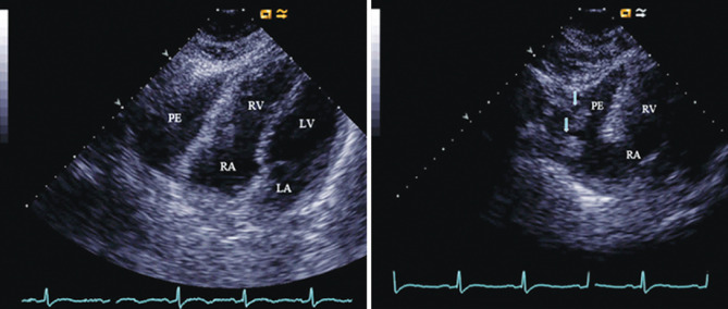
TTE subcostal view, left: shows moderate encysted anterior pericardial effusion, and right: zoom frame with multiple clots (arrows) in the pericardial space. RV: Right ventricle, RA: Right atrium, LV: Left ventricle, LA: Left atrium
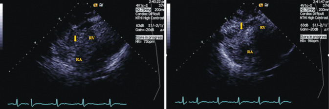
Agitated saline contrast injection: Left panel; starting to fill the right side, and right panel is a few seconds later image showing when contrast is cleared from the right side with some residual in the pericardial space (arrow), diagnostic of a connection between the right side and the pericardial space (pseudo-aneurysm). RV: Right ventricle, RA: Right atrium, LV: Left ventricle and LA: Left atrium
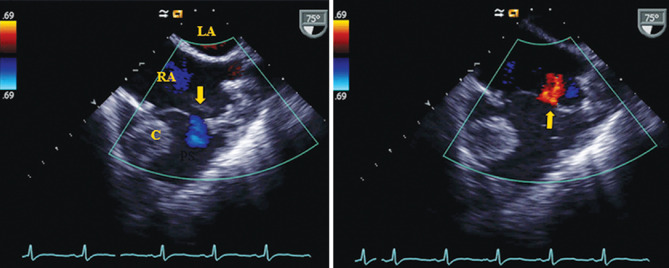
TEE showing right atrial pseudoaneurysm, and color flow saline into the pseudoaneurysm, left panel (arrow) and back into the RA, right panel (arrow). A clot (c) visualized in the pericardial space. RV: Right ventricle, RA: Right atrium, LV: Left ventricle and LA: Left atrium
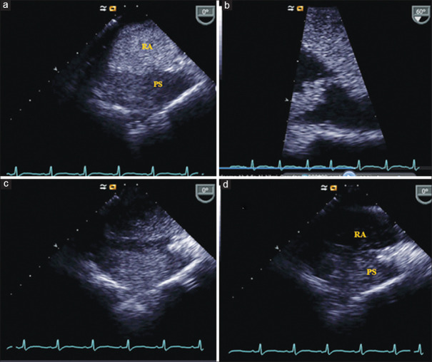
(a) TEE view showing agitated contrast saline starting to fill the RA and the pericardial space; (b) Zoom on the RA wall tear with the contrast passing through it into the pericardial space; (c) A few seconds later with the contrast in both the RA and the pericardial space, and (d) Later images showing the contrast is cleared from the RA side with a residual in the pericardial space, diagnostic of a pseudoaneurysm. RA: Right atrium, PS: Pericardial space
References
-
- Kitamura H, Okabayashi H, Hanyu M, Soga Y, Nomoto T, Johno H, et al. Successful enucleation of a giant cardiac hemangioendothelioma showing an unusual proliferation pattern. J Thorac Cardiovasc Surg. 2005;130:1199–201. - PubMed
-
- Gengenbach S, Ridker PM. Left ventricular hemangioma in Kasabach-Merritt syndrome. Am Heart J. 1991;121:202–3. - PubMed
-
- Marchiano D, Fisher F, Hofstetter S. Epithelioid hemangioendothelioma of the heart with distant metastases. A case report and literature review. J Cardiovasc Surg (Torino) 1993;34:529–33. - PubMed
-
- Hulten EA, Blankstein R. Pseudoaneurysms of the heart. Circulation. 2012;125:1920–5. - PubMed
Publication types
LinkOut - more resources
Full Text Sources