

Structure elucidation of the redox cofactor mycofactocin reveals oligo-glycosylation by MftF
- PMID: 33014324
- PMCID: PMC7491314
- DOI: 10.1039/d0sc01172j
Structure elucidation of the redox cofactor mycofactocin reveals oligo-glycosylation by MftF
Abstract
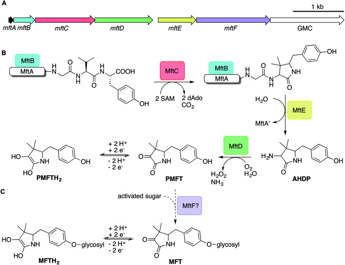
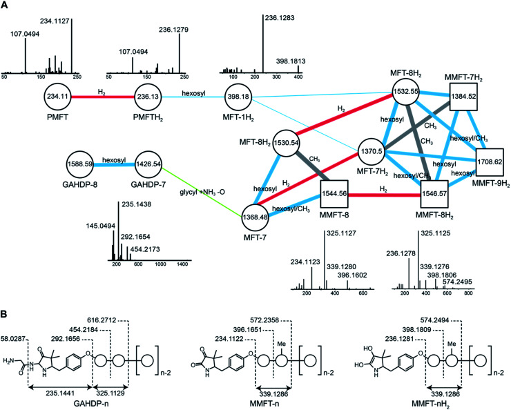
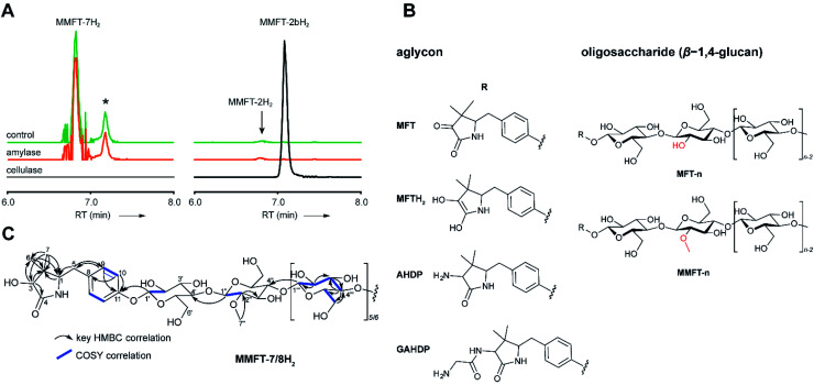
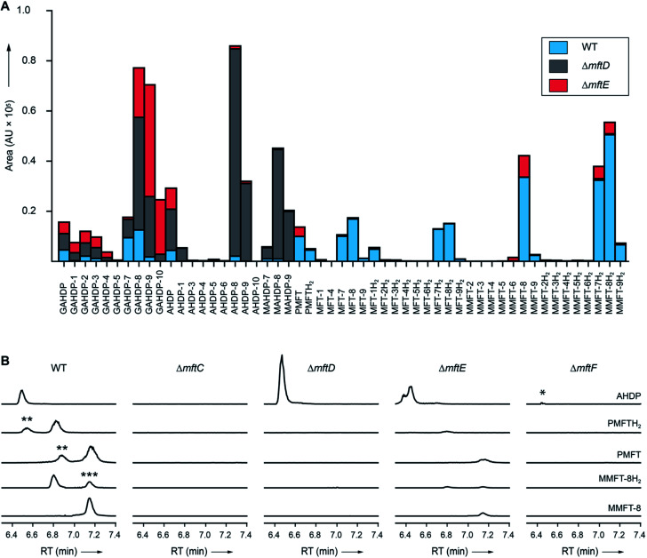
Mycofactocin (MFT) is a redox cofactor belonging to the family of ribosomally synthesized and post-translationally modified peptides (RiPPs) and is involved in alcohol metabolism of mycobacteria including Mycobacterium tuberculosis. A preliminary biosynthetic model had been established by bioinformatics and in vitro studies, while the structure of natural MFT and key biosynthetic steps remained elusive. Here, we report the discovery of glycosylated MFT by 13C-labeling metabolomics and establish a model of its biosynthesis in Mycolicibacterium smegmatis. Extensive structure elucidation including NMR revealed that MFT is decorated with up to nine β-1,4-linked glucose residues including 2-O-methylglucose. Dissection of biosynthetic genes demonstrated that the oligoglycosylation is catalyzed by the glycosyltransferase MftF. Furthermore, we confirm the redox cofactor function of glycosylated MFTs by activity-based metabolic profiling using the carveol dehydrogenase LimC and show that the MFT pool expands during cultivation on ethanol. Our results will guide future studies into the biochemical functions and physiological roles of MFT in bacteria.
This journal is © The Royal Society of Chemistry 2020.
Figures
Similar articles
-
Impact of Oxygen Supply and Scale Up on Mycobacterium smegmatis Cultivation and Mycofactocin Formation.Front Bioeng Biotechnol. 2020 Dec 3;8:593781. doi: 10.3389/fbioe.2020.593781. eCollection 2020. Front Bioeng Biotechnol. 2020. PMID: 33344432 Free PMC article.
-
Mycofactocin Is Associated with Ethanol Metabolism in Mycobacteria.mBio. 2019 May 21;10(3):e00190-19. doi: 10.1128/mBio.00190-19. mBio. 2019. PMID: 31113891 Free PMC article.
-
MftD Catalyzes the Formation of a Biologically Active Redox Center in the Biosynthesis of the Ribosomally Synthesized and Post-translationally Modified Redox Cofactor Mycofactocin.J Am Chem Soc. 2019 Aug 28;141(34):13582-13591. doi: 10.1021/jacs.9b06102. Epub 2019 Aug 15. J Am Chem Soc. 2019. PMID: 31381312 Free PMC article.
-
Occurrence, function, and biosynthesis of mycofactocin.Appl Microbiol Biotechnol. 2019 Apr;103(7):2903-2912. doi: 10.1007/s00253-019-09684-4. Epub 2019 Feb 18. Appl Microbiol Biotechnol. 2019. PMID: 30778644 Free PMC article. Review.
-
New Insights into the Biosynthetic Logic of Ribosomally Synthesized and Post-translationally Modified Peptide Natural Products.Cell Chem Biol. 2016 Jan 21;23(1):31-44. doi: 10.1016/j.chembiol.2015.11.012. Cell Chem Biol. 2016. PMID: 26933734 Free PMC article. Review.
Cited by
-
Transcriptomic Analysis of the Dual Response of Rhodococcus aetherivorans BCP1 to Inorganic Arsenic Oxyanions.Appl Environ Microbiol. 2022 Apr 12;88(7):e0220921. doi: 10.1128/aem.02209-21. Epub 2022 Mar 21. Appl Environ Microbiol. 2022. PMID: 35311511 Free PMC article.
-
Role of Premycofactocin Synthase in Growth, Microaerophilic Adaptation, and Metabolism of Mycobacterium tuberculosis.mBio. 2021 Aug 31;12(4):e0166521. doi: 10.1128/mBio.01665-21. Epub 2021 Jul 27. mBio. 2021. PMID: 34311585 Free PMC article.
-
S-Adenosylmethionine (SAM)-Dependent Methyltransferase MftM is Responsible for Methylation of the Redox Cofactor Mycofactocin.ACS Chem Biol. 2022 Nov 18;17(11):3207-3217. doi: 10.1021/acschembio.2c00659. Epub 2022 Oct 26. ACS Chem Biol. 2022. PMID: 36288793 Free PMC article.
-
New developments in RiPP discovery, enzymology and engineering.Nat Prod Rep. 2021 Jan 1;38(1):130-239. doi: 10.1039/d0np00027b. Epub 2020 Sep 16. Nat Prod Rep. 2021. PMID: 32935693 Free PMC article. Review.
-
Impact of Oxygen Supply and Scale Up on Mycobacterium smegmatis Cultivation and Mycofactocin Formation.Front Bioeng Biotechnol. 2020 Dec 3;8:593781. doi: 10.3389/fbioe.2020.593781. eCollection 2020. Front Bioeng Biotechnol. 2020. PMID: 33344432 Free PMC article.
References
-
- Buchmeier N. A., Newton G. L., Koledin T., Fahey R. C. Mol. Microbiol. 2003;47:1723–1732. - PubMed
LinkOut - more resources
Full Text Sources