Six Visual Rating Scales as A Biomarker for Monitoring Atrophied Brain Volume in Parkinson's Disease
- PMID: 33014524
- PMCID: PMC7505277
- DOI: 10.14336/AD.2019.1103
Six Visual Rating Scales as A Biomarker for Monitoring Atrophied Brain Volume in Parkinson's Disease
Abstract
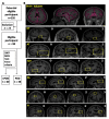
The focus of our investigation was to determine the feasibility of using six visual rating scales as whole-brain imaging markers for monitoring atrophied brain volume in Parkinson's disease (PD). This was a prospective cross-sectional single-center observational study. A total of 98 PD patients were enrolled and underwent an MRI scan and a battery of neuropsychological evaluations. The brain volume was calculated using the online resource MRICloud. Brain atrophy was rated based on six visual rating scales. Correlation analysis was performed between visual rating scores and brain volume and clinical features. We found a significant negative correlation between the total scores of visual rating scores and quantitative brain volume, indicating that six visual rating scales reliably reflect whole brain atrophy in PD. Multiple linear regression-based analyses indicated severer non-motor symptoms were significantly associated with higher scores on the visual rating scales. Furthermore, we performed sample size calculations to evaluate the superiority of visual rating scales; the result show that using total scores of visual rating scales as an outcome measure, sample sizes for differentiating cognition injury require significantly fewer subjects (n = 177) compared with using total brain volume (n = 2524). Our data support the use of the total visual rating scores rather than quantitative brain volume as a biomarker for monitoring cerebral atrophy.
Keywords: Parkinson’s disease; clinical trials; structural image; visual rating scale; whole brain atrophy.
copyright: © 2020 Lin et al.
Conflict of interest statement
Conflict of interest statement The authors do not have commercial or other associations that might pose a conflict of interest.
Figures


References
-
- Delgado-Alvarado M, Gago B, Navalpotro-Gomez I, Jimenez-Urbieta H, Rodriguez-Oroz MC (2016). Biomarkers for dementia and mild cognitive impairment in Parkinson’s disease. Mov Disord, 31:861-881. - PubMed
-
- Chetelat G, Baron JC (2003). Early diagnosis of Alzheimer’s disease: contribution of structural neuroimaging. Neuroimage, 18:525-541. - PubMed
-
- Davies RR, Scahill VL, Graham A, Williams GB, Graham KS, Hodges JR (2009). Development of an MRI rating scale for multiple brain regions: comparison with volumetrics and with voxel-based morphometry. Neuroradiology, 51:491-503. - PubMed
LinkOut - more resources
Full Text Sources
