Review
. 2020 Oct 1;11(5):1291-1316.
doi: 10.14336/AD.2019.1125. eCollection 2020 Oct.

Relationships between Mitochondrial Dysfunction and Neurotransmission Failure in Alzheimer's Disease

Affiliations
Review

Relationships between Mitochondrial Dysfunction and Neurotransmission Failure in Alzheimer's Disease

Kan Yin Wong et al. Aging Dis. .

Abstract

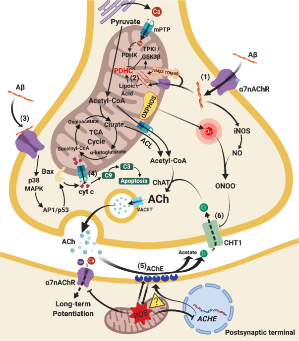
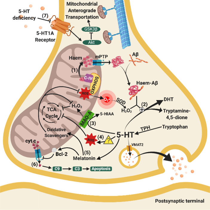
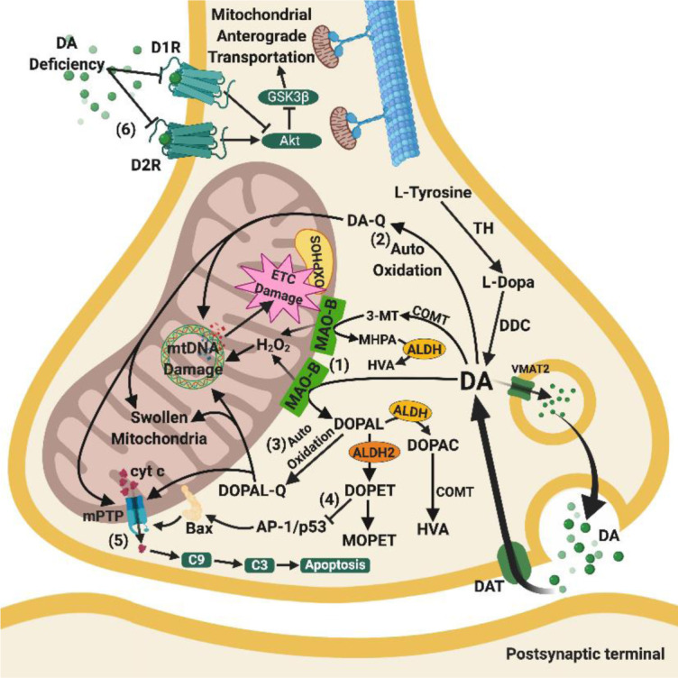
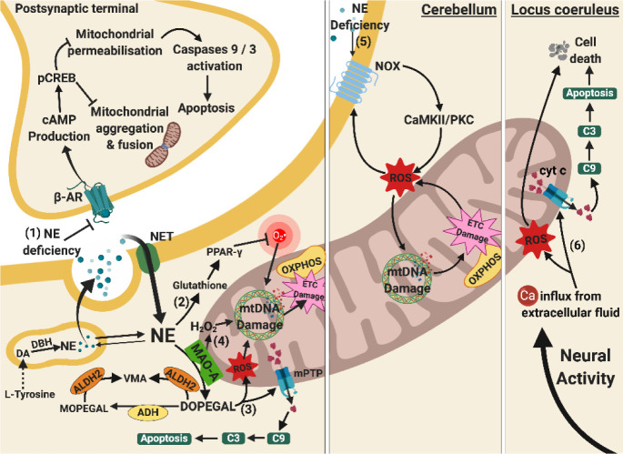
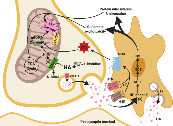
Besides extracellular deposition of amyloid beta and formation of phosphorylated tau in the brains of patients with Alzheimer's disease (AD), the pathogenesis of AD is also thought to involve mitochondrial dysfunctions and altered neurotransmission systems. However, none of these components can describe the diverse cognitive, behavioural, and psychiatric symptoms of AD without the pathologies interacting with one another. The purpose of this review is to understand the relationships between mitochondrial and neurotransmission dysfunctions in terms of (1) how mitochondrial alterations affect cholinergic and monoaminergic systems via disruption of energy metabolism, oxidative stress, and apoptosis; and (2) how different neurotransmission systems drive mitochondrial dysfunction via increasing amyloid beta internalisation, oxidative stress, disruption of mitochondrial permeabilisation, and mitochondrial trafficking. All these interactions are separately discussed in terms of neurotransmission systems. The association of mitochondrial dysfunctions with alterations in dopamine, norepinephrine, and histamine is the prospective goal in this research field. By unfolding the complex interactions surrounding mitochondrial dysfunction in AD, we can better develop potential treatments to delay, prevent, or cure this devastating disease.

Keywords: Alzheimer’s disease; mitochondrial dysfunction; monoaminergic; neurotransmission dysfunction.


Conflict of interest statement

Declaration of Competing Interest None.

Figures

Figure 1.
Figure 1.
Relationships between mitochondrial and cholinergic dysfunctions. (1) Upregulated α7-nAChRs on cellular and mitochondrial membrane internalise Aβ from the extracellular fluid to the cytosol and intramitochondrial matrix. The cytosolic Aβ induces iNOS production. (2) PDHC is inhibited by Aβ via activation of TPK1/GSK3β and inhibition of lipoic acid and by excess Ca2+ influx via activation of PDHK, leading to decreased acetyl-CoA and subsequent ACh synthesis. (3) Upregulated cellular α7-nAChRs combine with Aβ to stimulate p38 MAPK and AP1/p53 signalling pathways. Bax proteins on the mitochondrial membrane are activated and enhance mPTP opening. (4) The mPTP opening on unstable mitochondrial membrane leads to release of cyt c, which activates caspase 9/3-dependent apoptosis. (5) AChE activity is enhanced by oxidative stress from mitochondrial dysfunction possibly through inhibition of CHAT nuclear gene. The enhanced AChE activity produces higher ROS levels which damage mitochondria, forming a vicious cycle of lowered ACh levels in the synapse (further investigations are needed). (6) CHT1 is inhibited due to ONOO- produced by O2-• from mitochondria and NO from iNOS. Chloride ions reuptake decreases and reduces Ach synthesis. Arrows indicate stimulation, whereas a line with an end bar indicates inhibition. Dotted lines refer to inhibited pathways and question mark in a triangle () represents the need for future studies. Abbreviations: Acetyl-CoA, acetyl coenzyme A; ACh, acetylcholine; AChE, acetylcholinesterase; ACHE, gene that encodes acetylcholinesterase; ACL, ATP citrate lyase; AP-1/p53, activating protein-1 transcription factor / tumour protein p53; Aβ, amyloid-beta; Bax, Bcl-2-associated X protein; C3, caspase 3; C9, caspase 9; Ca, calcium ions; ChAT, choline acetyltransferase; CHT1, choline transporter 1; Cl, chlorine ions; cyt c, cytochrome c; iNOS, induced nitric oxide synthase; p38 MAPK, p38 mitogen-activated protein kinase signalling cascade; mPTP, mitochondrial permeability transition pore; Na, sodium ions; NO, nitric oxide; O2-•, superoxide radical; ONOO-, peroxynitrite; OXPHOS, oxidative phosphorylation; PDHC, pyruvate dehydrogenase complex; PDHK, pyruvate dehydrogenase kinase; ROS, reactive oxygen species; TCA cycle, tricarboxylic acid cycle; TIM23, translocase of the inner membrane 23; TOM40, translocase of outer mitochondrial membrane 40 homolog; TPKI/GSK3β, tau protein kinase I / Glycogen synthase kinase-3 beta signalling cascade; VAChT, vesicular acetylcholine transporter; α7nAChR, alpha 7 nicotinic acetylcholine receptor.
Figure 2.
Figure 2.
Relationships between mitochondrial and serotoninergic dysfunctions. (1) Haem from C-IV of OXPHOS released from mitochondria through increased mPTP opening bind with cytosol Aβ to form haem-Aβ complexes. (2) Mitochondrial dysfunction enhances the clearance of 5-HT through the combined effects of haem-Aβ complexes and peroxide activity (H2O2) from defective ETC. (3) Enhanced activity of MAO-A excessively breaks down 5-HT, leading to 5-HT-deficiency and H2O2, which lowers the efficiency of the TCA cycle. (4) 5-HT is associated with increased ROS levels (further research is needed to elucidate the precise molecular mechanisms), which damage enzymes in the TCA cycle. (5) Loss of 5-HT leads to the loss of anti-oxidative and anti-inflammatory melatonin, indirectly facilitating oxidative damage in mitochondria, reducing the activation of the Bcl-2 pathway. (6) Increased mPTP opening on the mitochondrial membrane leads to the release of cyt c, activating mitochondrial-mediated caspase-activated apoptosis. (7) Decreased 5-HT binding to 5HT-1A receptors hinders mitochondrial anterograde trafficking via inhibition of Akt and subsequent GSK3β stimulation, leading to altered normal energy distribution in the brain. Arrows indicate stimulation, whereas a line with an end bar indicates inhibition. A plus sign in circle (?) refers to catalysation and a question mark in triangle () represents the need for future studies. Abbreviations: 5-HIAA, 5-Hydroxyindoleacetic acid; 5-HT, serotonin; 5-HT1A receptor, serotonin 1A receptor; Akt, protein kinase B; Aβ, amyloid-beta; Bcl-2, B-cell lymphoma 2; C-IV, complex IV in electron transport chain; C3, caspase 3; C9, caspase 9; cyt c, cytochrome c; DHT, 5,7-dihydroxytryptamine; GSK3β, glycogen synthase kinase-3 beta; H2O2, hydrogen peroxide; MAO-A, monoamine oxidase A; mPTP, mitochondrial permeability transition pore; O2•-, superoxide radicals; OXPHOS, oxidative phosphorylation; ROS, reactive oxygen species; SOD, superoxide dismutase; TCA cycle, tricarboxylic acid cycle; TPH, tryptophan hydroxylase; VMAT2, vesicular monoamine transporter 2.
Figure 3.
Figure 3.
Relationships between mitochondrial and dopaminergic dysfunctions. (1) Overproduction of H2O2from MAO-B is considered a major peroxidative stressor that damages mtDNA and respiratory complexes of OXPHOS. (2) & (3) DA and DOPAL are auto-oxidised to their respective quinones by ROS formed from OXPHOS. The quinones aggravate the oxidative stress resulting in swollen mitochondria and nuclear DNA damage, and subsequent deficits in OXPHOS complexes. (4) Loss of ALDH2 on mitochondrial surface in AD leads to lower levels of DOPET, which is an anti-apoptotic metabolite cleaved from DOPAL. Loss of DOPET compromises the inhibition of AP-1/P53 and subsequent Bax activation and mPTP opening. (5) DA deficiency also contributes to the inactivation of dopamine 1 and 2 receptors, which control the anterograde transportation of mitochondria. However, investigations are required to elucidate how this loss relates to mitochondrial distribution in the brain. Arrows indicate stimulation, whereas a line with an end bar indicates inhibition. Abbreviations: 3-MT, 3-methoxy-4-hydroxyphenethylamine; Akt, protein kinase B; ALDH, aldehyde dehydrogenases; ALDH2, aldehyde dehydrogenase 2; AP-1/p53, activating protein-1 transcription factor / tumour protein p53; Bax, Bcl-2-associated X protein; C3, caspase3; C9, caspase 9; COMT, catechol-O-methyltransferase; cyt c, cytochrome c; D1R, dopamine 1 receptor; D2R, dopamine 2 receptor; DA, dopamine; DA-Q, dopamine quinone; DAT, dopamine transporter; DDC, DOPA decarboxylase; DOPA, dihydroxyphenylalanine; DOPAC, 3,4-dihydroxyphenylacetic acid; DOPAL, 3,4-dihydroxyphenylacetaldehyde; DOPAL-Q, 3,4-dihydroxyphenylacetaldehyde quinone; DOPET, 3,4-dihydroxyphenylethylamine; ETC, electron transport chain; GSK3β, glycogen synthase kinase-3 beta; H2O2, hydrogen peroxide; HVA, homovanillic acid; MAO-B, monoamine oxidase B; MHPA, 3-methoxy-4-hydroxyphenylacetaldehyde; MOPET, 4-(2-hydroxyethyl)-2-methoxyphenol; mPTP, mitochondrial permeability transition pore; mtDNA, mitochondrial deoxyribonucleic acid; OXPHOS, oxidative phosphorylation; TH, tyrosine hydroxylase; VMAT2, vesicular monoamine transporter 2.
Figure 4.
Figure 4.
Relationships between mitochondrial and norepinephrine dysfunctions. (1) NE deficiency leads to loss of its protective effects on postsynaptic neurons. Normally, β-AR activation leads to cAMP and pCREB production, reducing mitochondrial aggregation, fission, and membrane permeabilisation. These prevent mitochondrial morphology changes and mitochondrial-mediated caspases-activated apoptosis. (2) NE deficiency at presynaptic terminals lowers the level of glutathione and PPAR-γ activation, increasing O2•- via a receptor-independent pathway and predisposes to mtDNA damage. (3) DOPEGAL accumulates due to undermined ADH and ALDH2, generating both oxidative stress and mPTP opening. The mPTP opening facilitates release of pro-apoptotic cyt c and subsequent apoptosis. (4) Enhanced activity of MAO-A causes overproduction of H2O2 and along with oxidative stress from DOPEGAL stimulates mtDNA damage and ETC damage. (5) NE deficiency in cerebellum also causes mtDNA damage through the activation of NOX, CAMKII/PKCα signalling cascade, and ROS production. (6) Apoptosis of NE neurones at the locus coeruleus is associated with oxidative stress and mitochondrial-mediated caspases-dependent apoptosis due to Ca2+ influx upon neuronal activation, leading to reduced NE production. Arrows indicate stimulation, whereas a line with an end bar indicates inhibition. Dotted line represents a series of biochemical reactions. Abbreviations: ADH, alcohol dehydrogenase; ALDH 2, aldehyde dehydrogenase 2; C3, caspase3; C9, caspase 9; Ca, calcium ions; CAMKII/PKC, calmodulin-dependent protein kinase II/protein kinase C signalling cascade; cAMP, cyclic AMP; cyt c, cytochrome c; DA, dopamine; DBH, dopamine beta-hydroxylase; DOPEGAL, 3,4-dihydroxyphenylglycolaldehyde; ETC, electron transport chain; H2O2, hydrogen peroxide; MAO-A, monoamine oxidase A; MOPEGAL, 3-methoxy-4-hydroxyphenylglycolaldehyde; mPTP, mitochondrial permeability transitional pores; mtDNA, mitochondrial deoxyribonucleic acid; NE, norepinephrine; NET, norepinephrine transporter; NOX, NADPH oxidase; O2•-, superoxide radicals; OXPHOS, oxidative phosphorylation; pCREB, phosphorylated cyclic AMP response element binding; PPAR-γ, peroxisome proliferator-activated receptor gamma; ROS, reactive oxygen species; VMA, vanillylmandelic acid; β-AR, beta-2 adrenergic receptor.
Figure 5.
Figure 5.
Relationships between mitochondrial and histaminergic dysfunctions. (1) Histamine content in the synaptic cleft increases due to microglial release. (2) Increased HA concentration stimulates H1R and H4R, and subsequent NADPH oxidase, which enhances ROS production and leads to mtDNA damage and ETC damage. (3) Increased HA content at the synapse also induces the NF kappa-B/AP-1 signalling pathway facilitating iNOS synthesis of NO, which in turn leads to glutamate excitotoxicity, nitrosylation and nitrosation of proteins, particularly in OXPHOS (ETC damage) and TCA cycle. (4) Excessive H2O2produced from upregulated MAO-B leads to damage in mtDNA and the TCA cycle. These mechanisms ultimately damage mitochondrial bioenergy production in the brain. Arrows indicate stimulation, whereas a line with an end bar indicates inhibition. Abbreviations: AP-1, activating protein-1 transcription factor; ETC, electron transport chain; H1R, histaminergic 1 receptor; H2O2, hydrogen peroxide; H4R, histaminergic 4 receptor; HA, histamine; HDC, histidine decarboxylase; iNOS, induced nitric oxide synthase; MAO-B, monoamine oxidase B; mtDNA, mitochondrial deoxyribonucleic acid; N-MIAA, N-methyl indole acetic acid; NF-kappaB, nuclear factor kappa-light-chain-enhancer of activated B cells; NO, nitric oxide; NOX, NADPH oxidase; OXPHOS, oxidative phosphorylation; ROS, reactive oxygen species; TCA cycle, tricarboxylic acid cycle; VMAT2, vesicular monoamine transporter 2.

Similar articles

Cited by

References

    1. Cerejeira J, Lagarto L, Mukaetova-Ladinska EB (2012). Behavioral and psychological symptoms of dementia. Front Neurol, 3:73. - PMC - PubMed
    1. Alzheimer’s Association (2018). 2018 Alzheimer’s disease facts and figures. Alzheimer’s & Dementia, 14:367-429.
    1. Prince M, Bryce R, Albanese E, Wimo A, Ribeiro W, Ferri CP (2013). The global prevalence of dementia: A systematic review and metaanalysis. Alzheimer’s & Dementia, 9:63-75.e62. - PubMed
    1. Hardy JA, Higgins GA (1992). Alzheimer’s disease: the amyloid cascade hypothesis. Science, 256:184-185. - PubMed
    1. Querfurth HW, LaFerla FM (2010). Alzheimer’s disease. N Engl J Med, 362:329-344. - PubMed

LinkOut - more resources