Review
. 2020 Oct 1;11(5):1317-1328.
doi: 10.14336/AD.2019.1116. eCollection 2020 Oct.

Infrapatellar Fat Pad and Knee Osteoarthritis

Affiliations
Review

Infrapatellar Fat Pad and Knee Osteoarthritis

Ni Zeng et al. Aging Dis. .

Abstract

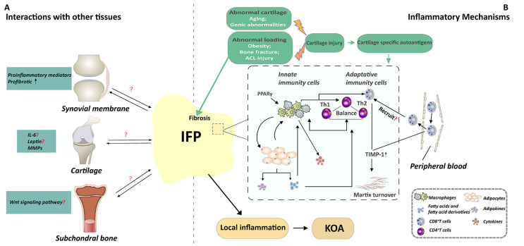
Osteoarthritis is the most prevalent arthritis typically characterized by degradation of cartilage. However, its pathogenesis is not fully understood. Currently, osteoarthritis is best considered a disease of the whole "joint organ". Infrapatellar fat pad (IFP), an adipose tissue near synovium, is now attaching importance to researchers for its inflammatory phenotype. In this narrative review, a large body of evidence has been gathered for the involvement of IFP in the development of knee osteoarthritis. Additionally, the underlying mechanisms of how IFP can be involved in this process have been proposed. However, further investigations are needed to better understand its precise role in this process and its underlying mechanism, and beyond that, to develop new strategies to slow down the degenerative process and explore an effective and timely diagnosis of the disease.

Keywords: crosstalk; infrapatellar fat pad; knee osteoarthritis; local inflammation.


Conflict of interest statement

Competing interests The authors declare that they have no competing interests.

Figures

Figure 1.
Figure 1.
A mechanistic model depicting how IFP is involved in OA pathogenesis. It is supposed that the IFP may be involved in the local inflammation state and/or that it might interact with other periarticular tissues, thus contributing to the development of KOA. (A). IFP may interact with cartilage, synovial membrane and subchondral bone. IFP may interact with the synovial tissue through affecting the inflammatory response and promoting the fibrosis of the synovial cells. In parallel, IFP plays either a deleterious or a protective role on cartilage by altering the expression of IL-6 or leptin or MMPs. While the influence of IFP on subchondral bone has not yet been investigated clear, IFP may interact with subchondral bone through Wnt signaling pathway. Reciprocally, these joint tissues could also modulate the IFP. (B). Many factors such as obesity, bone fracture, and aging are related to abnormal loading or abnormal cartilage, and eventually lead to cartilage injury. Then, the immune response of IFP may be activated by the cartilage specific autoantigens. The immunophenotype of immune cells of IFP is related to inflammatory phenotype of IFP, which is the potential mechanisms of the involvement of IFP in the development of OA. Macrophages consist of is type M1 and M2 macrophage. PPARγ-regulated genes are upregulated in M2 macrophage. Macrophages can interact with T cells and adipocytes to participate in local inflammation state. First, CD4 ?T cell also called Th cells, it contains two subtypes named Th1 and Th2. The balance between type Th1 and Th2 type signals may affect the recruitment and activation of macrophages in the adipose tissue, resulting in pathogenic and inflammatory conditions or non-inflammatory and protective environments. Then, adipocytes secrete fatty acids and fatty acid derivatives which are highly immune-regulated, to modulate the phenotype of macrophages and CD4+T cells. Moreover, CD8+ T present a higher level of activation in the IFP, and the activated CD8+ T cells in IFP may recruited into the tissues by preactivated circulating peripheral blood. In addition, CD8+T cells may modulate matrix turnover, and thus contribute to the development of OA by up-regulating TIMP-1. These activated immune cells and adipocytes alter the inflammatory phenotype of IFP, participate in the local inflammation of the whole joint, and ultimately lead to KOA. Short title of the first figure. The figure caption should begin with a title (an overall descriptive statement of the figure) followed by additional text. The legends should be placed immediately after each figure.

Similar articles

Cited by

References

    1. Favero M, El-Hadi H, Belluzzi E, Granzotto M, Porzionato A, Sarasin G, et al. (2017). Infrapatellar fat pad features in osteoarthritis: a histopathological and molecular study. Rheumatology (Oxford), 56:1784-1793. - PubMed
    1. Pan F, Han W, Wang X, Liu Z, Jin X, Antony B, et al. (2015). A longitudinal study of the association between infrapatellar fat pad maximal area and changes in knee symptoms and structure in older adults. Ann Rheum Dis, 74:1818-1824. - PubMed
    1. Ioan-Facsinay A, Kloppenburg M (2013). An emerging player in knee osteoarthritis: the infrapatellar fat pad. Arthritis Res Ther, 15:225. - PMC - PubMed
    1. Bos SD, Slagboom PE, Meulenbelt I (2008). New insights into osteoarthritis: early developmental features of an ageing-related disease. Curr Opin Rheumatol, 20:553-559. - PubMed
    1. Zhu Z, Otahal P, Wang B, Jin X, Laslett LL, Wluka AE, et al. (2017). Cross-sectional and longitudinal associations between serum inflammatory cytokines and knee bone marrow lesions in patients with knee osteoarthritis. Osteoarthritis Cartilage, 25:499-505. - PubMed

LinkOut - more resources