

Registration of fluorescein angiography and optical coherence tomography images of curved retina via scanning laser ophthalmoscopy photographs
- PMID: 33014544
- PMCID: PMC7510895
- DOI: 10.1364/BOE.395784
Registration of fluorescein angiography and optical coherence tomography images of curved retina via scanning laser ophthalmoscopy photographs
Abstract
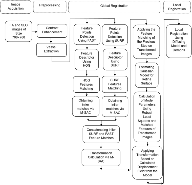
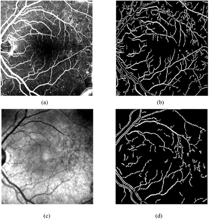
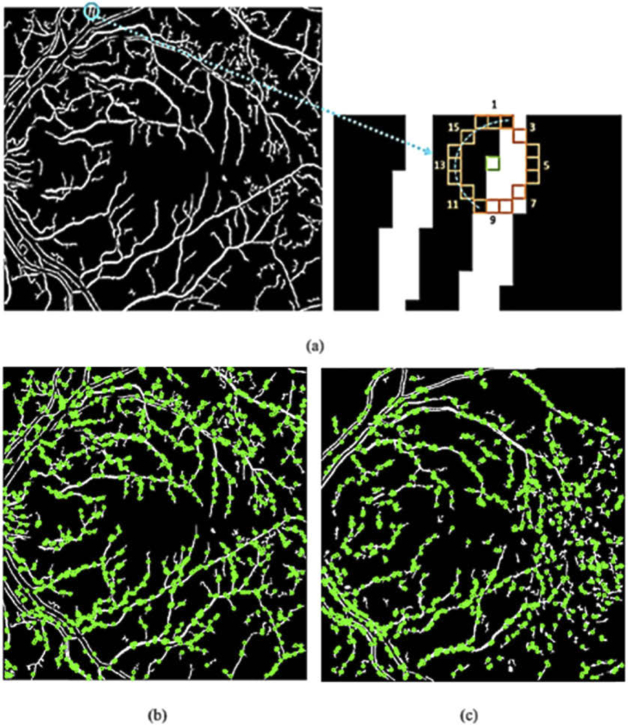
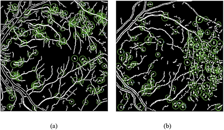
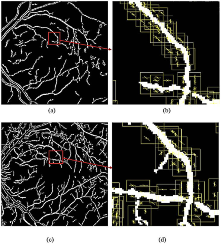
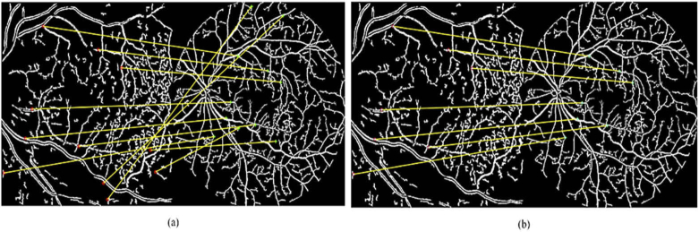
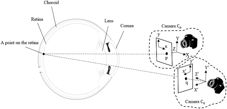
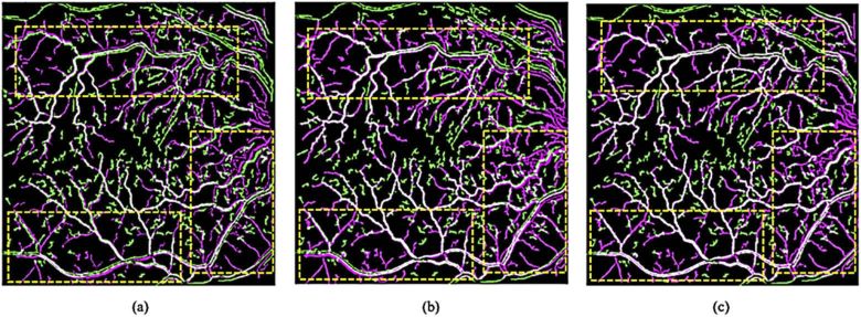
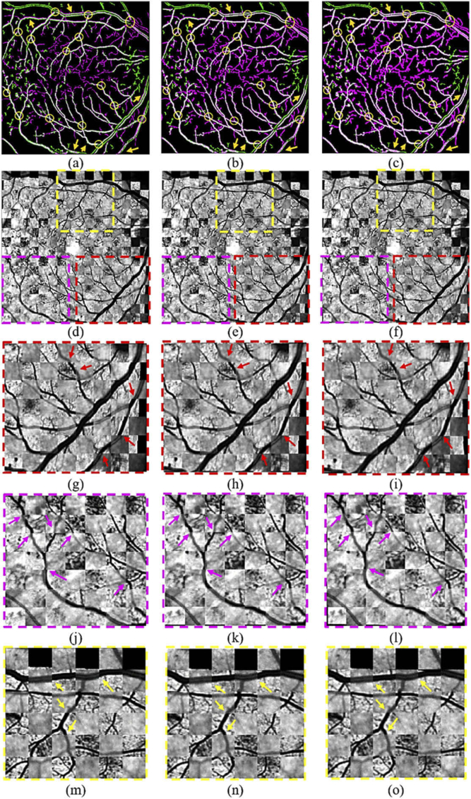
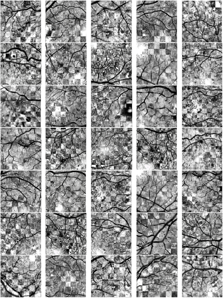
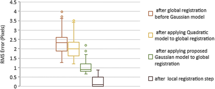
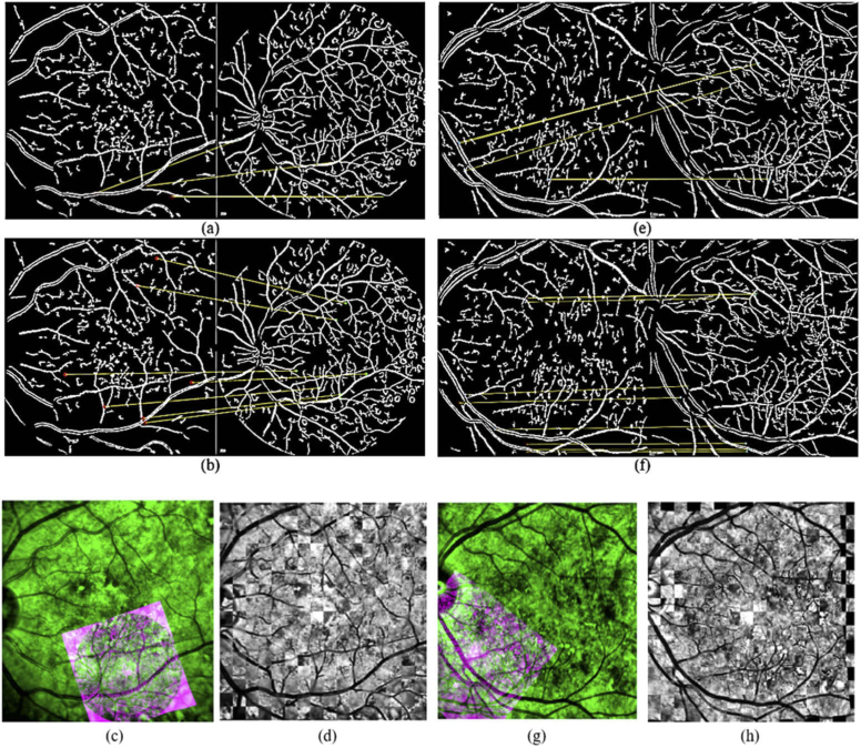
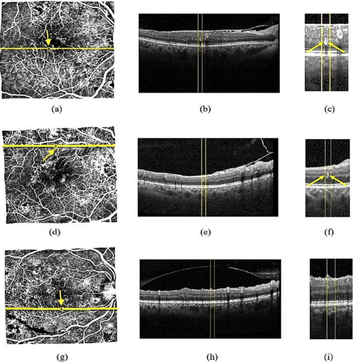
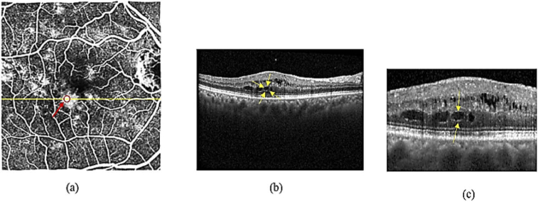
Accurate and automatic registration of multimodal retinal images such as fluorescein angiography (FA) and optical coherence tomography (OCT) enables utilization of supplementary information. FA is a gold standard imaging modality that depicts neurovascular structure of retina and is used for diagnosing neurovascular-related diseases such as diabetic retinopathy (DR). Unlike FA, OCT is non-invasive retinal imaging modality that provides cross-sectional data of retina. Due to differences in contrast, resolution and brightness of multimodal retinal images, the images resulted from vessel extraction of image pairs are not exactly the same. Also, prevalent feature detection, extraction and matching schemes do not result in perfect matches. In addition, the relationships between retinal image pairs are usually modeled by affine transformation, which cannot generate accurate alignments due to the non-planar retina surface. In this paper, a precise registration scheme is proposed to align FA and OCT images via scanning laser ophthalmoscopy (SLO) photographs as intermediate images. For this purpose, first a retinal vessel segmentation is applied to extract main blood vessels from the FA and SLO images. Next, a novel global registration is proposed based on the Gaussian model for curved surface of retina. For doing so, first a global rigid transformation is applied to FA vessel-map image using a new feature-based method to align it with SLO vessel-map photograph, in a way that outlier matched features resulted from not-perfect vessel segmentation are completely eliminated. After that, the transformed image is globally registered again considering Gaussian model for curved surface of retina to improve the precision of the previous step. Eventually a local non-rigid transformation is exploited to register two images perfectly. The experimental results indicate the presented scheme is more precise compared to other registration methods.
© 2020 Optical Society of America under the terms of the OSA Open Access Publishing Agreement.
Conflict of interest statement
The authors declare that there are no conflicts of interest related to this article.
Figures
References
-
- Khaderi K., Ahmed K., Berry J., Labriola L., Cornwell R., “Retinal imaging modalities: advantages and limitations for clinical practice,” Retinal Physician 8 (2011).
-
- Wang H., Chhablani J., Freeman W. R., Chan C. K., Kozak I., Bartsch D.-U., Cheng L., “Characterization of diabetic microaneurysms by simultaneous fluorescein angiography and spectral-domain optical coherence tomography,” Am. J. Ophthalmol. 153(5), 861–867.e1 (2012). 10.1016/j.ajo.2011.10.005 - DOI - PMC - PubMed
LinkOut - more resources
Full Text Sources