. 2020 Jun 22;11(7):3913-3926.
doi: 10.1364/BOE.394921. eCollection 2020 Jul 1.

Silicon photonics-based laser Doppler vibrometer array for carotid-femoral pulse wave velocity (PWV) measurement

Affiliations

Silicon photonics-based laser Doppler vibrometer array for carotid-femoral pulse wave velocity (PWV) measurement

Yanlu Li et al. Biomed Opt Express. .

Abstract

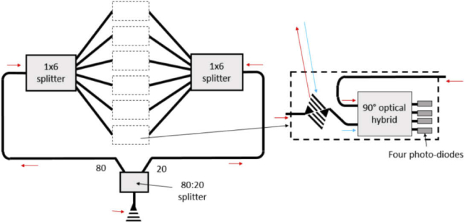
Pulse wave velocity (PWV) is a reference measure for aortic stiffness, itself an important biomarker of cardiovascular risk. To enable low-cost and easy-to-use PWV measurement devices that can be used in routine clinical practice, we have designed several handheld PWV sensors using miniaturized laser Doppler vibrometer (LDV) arrays in a silicon photonics platform. The LDV-based PWV sensor design and the signal processing protocol to obtain pulse transit time (PTT) and carotid-femoral PWV in a feasibility study in humans, are described in this paper. Compared with a commercial reference PWV measurement system, measuring arterial pressure waveforms by applanation tonometry, LDV-based displacement signals resulted in more complex signals. However, we have shown that it is possible to identify reliable fiducial points for PTT calculation using the maximum of the 2nd derivative algorithm in LDV-based signals, comparable to those obtained by the reference technique, applanation tonometry.


Conflict of interest statement

The authors declare no conflicts of interest.

Figures

Fig. 1.
Fig. 1.
(a) The configuration of the six-beam sensor head. (b) The three major parts of the PWV measurement system.
Fig. 2.
Fig. 2.
Different ways of using the two sensor heads. a. Detached. b. Attached without spacer. c.Attached with a spacer (13 mm). Note the sensor heads in the figures are non-functional 1:1 models with different colors from the working devices.
Fig. 3.
Fig. 3.
(a) The inside of the photonic head. PIC stands for photonic integrated circuit, MOB stands for micro-optical bench and BL stands for ball lens. (b) the use of the sensing spacers.
Fig. 4.
Fig. 4.
Schematic showing the PIC design with six LDVs.
Fig. 5.
Fig. 5.
(a) Schematic of the MOB design and a side-view microscope image of the MOB. (b) The mounting of the PIC with PCB.
Fig. 6.
Fig. 6.
Schematic show of the optical system. The sizes of the lenses and PIC are exaggerated in the figure to clearly show the configuration of the lens system.
Fig. 7.
Fig. 7.
(a) The target used to test the positions of the output beams. (b) the measured time delay vs set time delay for the time delay measurement (note the 5 ms sample is omitted from the plot to allow the fit of the shorter delay times to be more clearly seen).
Fig. 8.
Fig. 8.
(a) A picture of the measurement setting for carotid-femoral PWV. The two sensor heads of the LDV device are detached and positioned simultaneously on the carotid site (LDV1) and on the femoral site (LDV2). An ECG signal is simultaneously recorded. (b) Schematic representation of the 16 beam combinations for PTT calculation, arising by analysis of the 6 sensing beams from LDV1 sensor head, on the carotid site (beams 1.1 to 1.6) and the 6 sensing beams from LDV2 sensor head, on the femoral site (beams 2.1 to 2.6).
Fig. 9.
Fig. 9.
Example of the displacement LDV signals (left) and tonometric arterial pressure waveforms (right) obtained from carotid and femoral measurements by LDV (one beam shown per location) in a young (a), a middle aged (b) individual. The corresponding acceleration signals exhibit peaks in systole, after the ECG R-peak, which allows identification of the wave-foot in both LDV and tonometric signals. A time delay between the acceleration peaks of the carotid (blue) and femoral (red) is clearly visible.
Fig. 10.
Fig. 10.
Example of the displacement LDV signals (left) and tonometric arterial pressure waveforms (right) obtained from carotid and femoral measurements by LDV (one beam shown per location) in an old individual. A time delay between the acceleration peaks of the carotid (blue) and femoral (red) is clearly visible.
Fig. 11.
Fig. 11.
Agreement between PTT calculated by means of ECG-dependent and ECG-independent algorithms. a) linear regression, equation PTT (ECG-independent algorithm) = 3.95 ± 0.96 × PTT (ECG-dependent algorithm). b) Bland Altman Plot. The red line represents the mean difference between PTT calculated by the ECG-independent and the ECG-dependent algorithm, whereas the blue lines represent its 95% confidence limits.

References

    1. van Sloten T., Sedaghat S., Laurent S., London G., Pannier B., Ikram M., Kavousi M., Mattace-Raso F., Franco O., Boutouyrie P., Stehouwer C., “Carotid stiffness is associated with incident stroke: a systematic review and individual participant data meta-analysis,” J. Am. Coll. Cardiol. 66(19), 2116–2125 (2015).10.1016/j.jacc.2015.08.888 - DOI - PubMed
    1. Nichols W., O’Rourke M., Mcdonald’s Blood Flow in Arteries: Theoretical, Experimental, and Clinical Principles, 4th ed (Edward Arnold, 1998
    1. The Reference Values for Arterial Stiffness’ Collaboration , “Determinants of pulse wave velocity in healthy people and in the presence of cardiovascular risk factors: ‘establishing normal and reference values’,” Eur. Heart J. 31(19), 2338–2350 (2010).10.1093/eurheartj/ehq165 - DOI - PMC - PubMed
    1. Ben-Shlomo Y., Spears M., Boustred C., May M., Anderson S., Benjamin E., Boutouyrie P., Cameron J., Chen C., Cruickshank J., Hwang S., Lakatta E., Laurent S., Maldonado J., Mitchell G., Najjar S., Newman A., Ohishi M., Pannier B., Pereira T., Vasan R., Shokawa T., Sutton-Tyrell K., Verbeke F., Wang K., Webb D., Willum Hansen T., Zoungas S., McEniery C., Cockcroft J., Wilkinson I., “Aortic pulse wave velocity improves cardiovascular event prediction: an individual participant meta-analysis of prospective observational data from 17,635 subjects,” J. Am. Coll. Cardiol. 63(7), 636–646 (2014).10.1016/j.jacc.2013.09.063 - DOI - PMC - PubMed
    1. Vlachopoulos C., Aznaouridis K., Stefanadis C., “Prediction of cardiovascular events and all-cause mortality with arterial stiffness: a systematic review and meta-analysis,” J. Am. Coll. Cardiol. 55(13), 1318–1327 (2010).10.1016/j.jacc.2009.10.061 - DOI - PubMed

LinkOut - more resources