

Moxifloxacin based axially swept wide-field fluorescence microscopy for high-speed imaging of conjunctival goblet cells
- PMID: 33014588
- PMCID: PMC7510874
- DOI: 10.1364/BOE.401896
Moxifloxacin based axially swept wide-field fluorescence microscopy for high-speed imaging of conjunctival goblet cells
Abstract
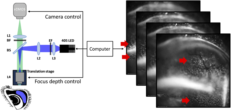
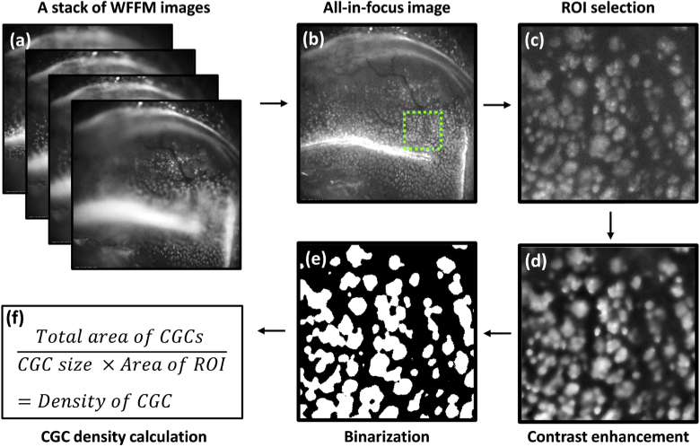
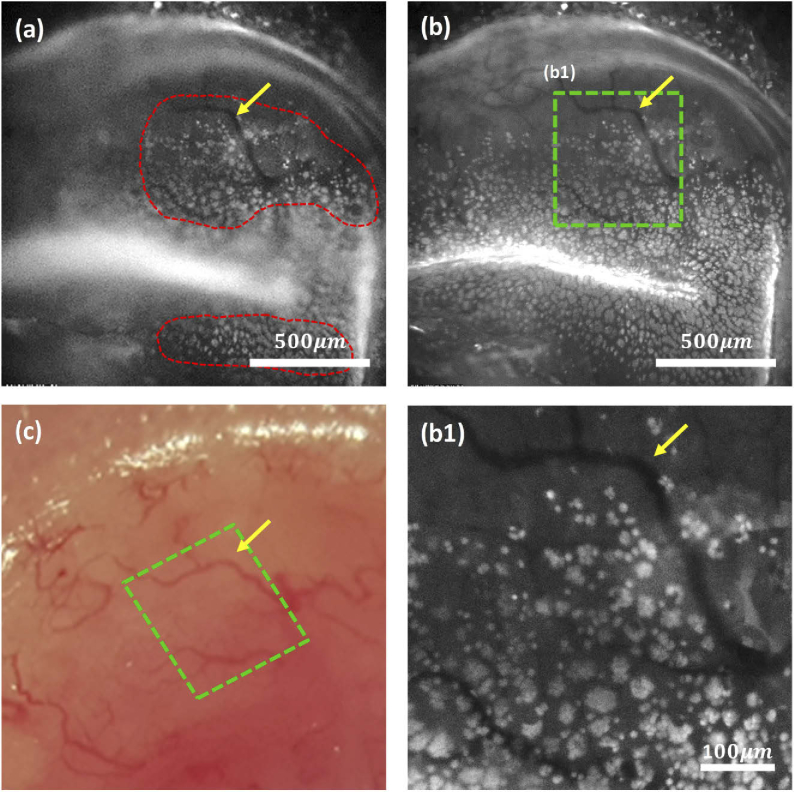
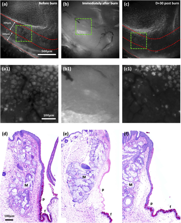
Goblet cells (GCs) in the conjunctiva are specialized epithelial cells producing mucins on the ocular surface. GCs play important roles in maintaining homeostasis of the ocular surface, and GC dysfunction is associated with various complications including dry eye diseases. Current GC examination methods, which are conjunctival impression cytology and confocal reflection microscopy, have limitations for routine examination. Fluorescence microscopy using moxifloxacin was recently introduced as a non-invasive and high-contrast imaging method, but further development is needed to be used for GC examination. Here we developed a non-invasive high-speed high-contrast GC imaging method, called moxifloxacin based axially swept wide-field fluorescence microscopy (MBAS-WFFM). This method acquired multiple fluorescence images with the axial sweeping of the focal plane to capture moxifloxacin labeled GCs on the tilted conjunctival surface in focus and generated all-in-focus images by combining the acquired images. The imaging field of view and imaging speed were increased to 1.6 mm × 1.6 mm and 30 fps. An image processing method was developed for the analysis of GC density. MBAS-WFFM was applied to alkali burn mouse models and detected GC damage and recovery via longitudinal imaging. MBAS-WFFM could assess the status of GCs rapidly and non-invasively. We anticipate MBAS-WFFM to be a starting point for non-invasive GC examination and the diagnosis of GC associated diseases. For example, MBAS-WFFM could be used to classify dry eye diseases into detail categories for effective treatment.
© 2020 Optical Society of America under the terms of the OSA Open Access Publishing Agreement.
Conflict of interest statement
SK, MJK, and KHK are authors of a patent filed for moxifloxacin-based imaging of goblet cells.
Figures
Similar articles
-
In Vivo Noncontact Imaging of Conjunctival Goblet Cells with Customized Widefield Fluorescence Microscopy.Ophthalmol Sci. 2025 Jan 12;5(3):100712. doi: 10.1016/j.xops.2025.100712. eCollection 2025 May-Jun. Ophthalmol Sci. 2025. PMID: 40130265 Free PMC article.
-
In vivo fluorescence imaging of conjunctival goblet cells.Sci Rep. 2019 Oct 29;9(1):15457. doi: 10.1038/s41598-019-51893-4. Sci Rep. 2019. PMID: 31664078 Free PMC article.
-
Moxifloxacin-Based Extended Depth-of-Field Fluorescence Microscopy for Real-Time Conjunctival Goblet Cell Examination.IEEE Trans Med Imaging. 2022 Aug;41(8):2004-2008. doi: 10.1109/TMI.2022.3151944. Epub 2022 Aug 1. IEEE Trans Med Imaging. 2022. PMID: 35167445
-
Conjunctival Goblet Cells, the Overlooked Cells in Glaucoma Treatment.J Glaucoma. 2019 Apr;28(4):325-333. doi: 10.1097/IJG.0000000000001168. J Glaucoma. 2019. PMID: 30585937 Review.
-
Secreted Mucins on the Ocular Surface.Invest Ophthalmol Vis Sci. 2018 Nov 1;59(14):DES151-DES156. doi: 10.1167/iovs.17-23623. Invest Ophthalmol Vis Sci. 2018. PMID: 30481820 Review.
Cited by
-
In Vivo Noncontact Imaging of Conjunctival Goblet Cells with Customized Widefield Fluorescence Microscopy.Ophthalmol Sci. 2025 Jan 12;5(3):100712. doi: 10.1016/j.xops.2025.100712. eCollection 2025 May-Jun. Ophthalmol Sci. 2025. PMID: 40130265 Free PMC article.
-
Noninvasive Imaging of Conjunctival Goblet Cells as a Method for Diagnosing Dry Eye Disease in an Experimental Mouse Model.Transl Vis Sci Technol. 2023 Dec 1;12(12):22. doi: 10.1167/tvst.12.12.22. Transl Vis Sci Technol. 2023. PMID: 38149964 Free PMC article.
-
Deep learning framework for automated goblet cell density analysis in in-vivo rabbit conjunctiva.Sci Rep. 2023 Dec 21;13(1):22839. doi: 10.1038/s41598-023-49275-y. Sci Rep. 2023. PMID: 38129447 Free PMC article.
-
Multi-Focus Image Fusion Using Focal Area Extraction in a Large Quantity of Microscopic Images.Sensors (Basel). 2021 Nov 5;21(21):7371. doi: 10.3390/s21217371. Sensors (Basel). 2021. PMID: 34770677 Free PMC article.
References
LinkOut - more resources
Full Text Sources
Miscellaneous