

Deep learning architecture "LightOCT" for diagnostic decision support using optical coherence tomography images of biological samples
- PMID: 33014597
- PMCID: PMC7510870
- DOI: 10.1364/BOE.395487
Deep learning architecture "LightOCT" for diagnostic decision support using optical coherence tomography images of biological samples
Abstract
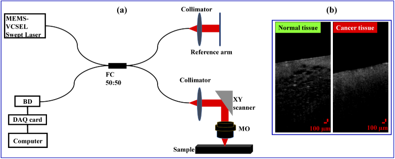
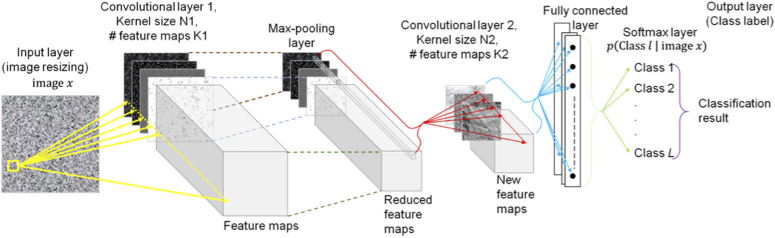
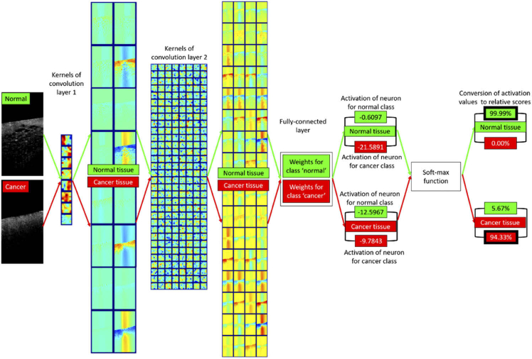
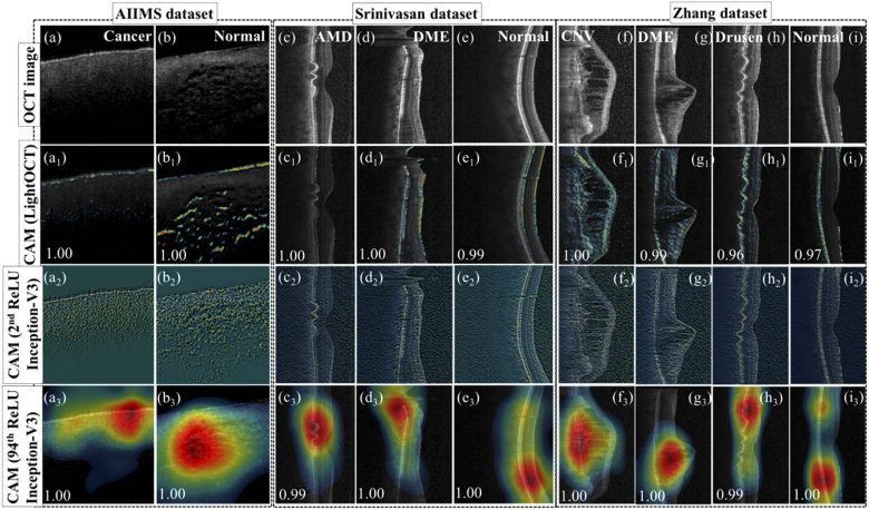
Optical coherence tomography (OCT) is being increasingly adopted as a label-free and non-invasive technique for biomedical applications such as cancer and ocular disease diagnosis. Diagnostic information for these tissues is manifest in textural and geometric features of the OCT images, which are used by human expertise to interpret and triage. However, it suffers delays due to the long process of the conventional diagnostic procedure and shortage of human expertise. Here, a custom deep learning architecture, LightOCT, is proposed for the classification of OCT images into diagnostically relevant classes. LightOCT is a convolutional neural network with only two convolutional layers and a fully connected layer, but it is shown to provide excellent training and test results for diverse OCT image datasets. We show that LightOCT provides 98.9% accuracy in classifying 44 normal and 44 malignant (invasive ductal carcinoma) breast tissue volumetric OCT images. Also, >96% accuracy in classifying public datasets of ocular OCT images as normal, age-related macular degeneration and diabetic macular edema. Additionally, we show ∼96% test accuracy for classifying retinal images as belonging to choroidal neovascularization, diabetic macular edema, drusen, and normal samples on a large public dataset of more than 100,000 images. The performance of the architecture is compared with transfer learning based deep neural networks. Through this, we show that LightOCT can provide significant diagnostic support for a variety of OCT images with sufficient training and minimal hyper-parameter tuning. The trained LightOCT networks for the three-classification problem will be released online to support transfer learning on other datasets.
© 2020 Optical Society of America under the terms of the OSA Open Access Publishing Agreement.
Conflict of interest statement
Authors declare no competing interest.
Figures

Similar articles
-
Fully automated detection of retinal disorders by image-based deep learning.Graefes Arch Clin Exp Ophthalmol. 2019 Mar;257(3):495-505. doi: 10.1007/s00417-018-04224-8. Epub 2019 Jan 4. Graefes Arch Clin Exp Ophthalmol. 2019. PMID: 30610422
-
Stitched vision transformer for age-related macular degeneration detection using retinal optical coherence tomography images.PLoS One. 2024 Jun 5;19(6):e0304943. doi: 10.1371/journal.pone.0304943. eCollection 2024. PLoS One. 2024. PMID: 38837967 Free PMC article.
-
Deep Learning Classification of Drusen, Choroidal Neovascularization, and Diabetic Macular Edema in Optical Coherence Tomography (OCT) Images.Cureus. 2023 Jul 9;15(7):e41615. doi: 10.7759/cureus.41615. eCollection 2023 Jul. Cureus. 2023. PMID: 37565126 Free PMC article.
-
Automated Segmentation and Quantification of Drusen in Fundus and Optical Coherence Tomography Images for Detection of ARMD.J Digit Imaging. 2018 Aug;31(4):464-476. doi: 10.1007/s10278-017-0038-7. J Digit Imaging. 2018. PMID: 29204763 Free PMC article. Review.
-
[Clinical applications of OCT angiography].Ophthalmologe. 2016 Jan;113(1):14-22. doi: 10.1007/s00347-015-0192-6. Ophthalmologe. 2016. PMID: 26694492 Review. German.
Cited by
-
Inflation of test accuracy due to data leakage in deep learning-based classification of OCT images.Sci Data. 2022 Sep 22;9(1):580. doi: 10.1038/s41597-022-01618-6. Sci Data. 2022. PMID: 36138025 Free PMC article.
-
Differentiation of breast tissue types for surgical margin assessment using machine learning and polarization-sensitive optical coherence tomography.Biomed Opt Express. 2021 Apr 29;12(5):3021-3036. doi: 10.1364/BOE.423026. eCollection 2021 May 1. Biomed Opt Express. 2021. PMID: 34168912 Free PMC article.
-
Multi-class classification of breast tissue using optical coherence tomography and attenuation imaging combined via deep learning.Biomed Opt Express. 2022 May 12;13(6):3380-3400. doi: 10.1364/BOE.455110. eCollection 2022 Jun 1. Biomed Opt Express. 2022. PMID: 35781967 Free PMC article.
-
Non-transfer Deep Learning of Optical Coherence Tomography for Post-hoc Explanation of Macular Disease Classification.Proc (IEEE Int Conf Healthc Inform). 2021 Aug;2021:48-52. doi: 10.1109/ichi52183.2021.00020. Epub 2021 Oct 15. Proc (IEEE Int Conf Healthc Inform). 2021. PMID: 36168324 Free PMC article.
-
Dense Convolutional Neural Network-Based Deep Learning Pipeline for Pre-Identification of Circular Leaf Spot Disease of Diospyros kaki Leaves Using Optical Coherence Tomography.Sensors (Basel). 2024 Aug 21;24(16):5398. doi: 10.3390/s24165398. Sensors (Basel). 2024. PMID: 39205092 Free PMC article.
References
-
- Butola A., Joshi T., Ahmad A., Dubey V., Senthilkumaran P., Mehta D. S., “3D topography and tomography of multilayered freeform optical surfaces using large-range measurement swept-source low-coherence interferometry,” Laser Phys. 28(11), 116101 (2018).10.1088/1555-6611/aad89e - DOI
-
- Nolan R. M., Adie S. G., Marjanovic M., Chaney E. J., South F. A., Monroy G. L., Shemonski N. D., Erickson-Bhatt S. J., Shelton R. L., Bower A. J., “Intraoperative optical coherence tomography for assessing human lymph nodes for metastatic cancer,” BMC Cancer 16(1), 144 (2016).10.1186/s12885-016-2194-4 - DOI - PMC - PubMed
LinkOut - more resources
Full Text Sources