. 2020 Sep 2:10:1483.
doi: 10.3389/fonc.2020.01483. eCollection 2020.

Mechanisms and Applications of the Anti-cancer Effect of Pharmacological Ascorbic Acid in Cervical Cancer Cells

Affiliations

Mechanisms and Applications of the Anti-cancer Effect of Pharmacological Ascorbic Acid in Cervical Cancer Cells

Tsai-Ming Wu et al. Front Oncol. .

Abstract

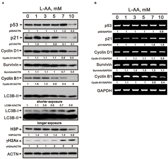
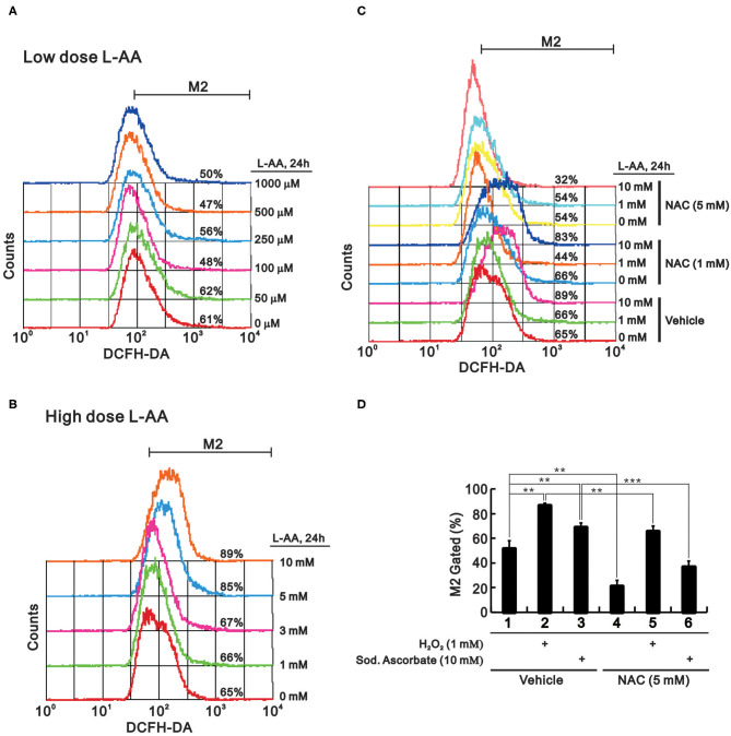
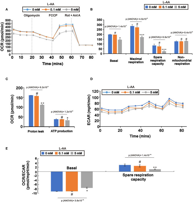
In recent years, L-ascorbic acid (L-AA), or vitamin C, has been attracting attention as a potential anticancer drug that mediates hydrogen peroxide-induced oxidation and ten-eleven translocation 2-catalyzed DNA demethylation. However, the precise mechanism by which L-AA acts remains unclear. We examined the cytotoxic effects of L-AA or sodium ascorbate in human cervical carcinoma cells by assessing cell viability, expression of cell cycle-related mRNAs and proteins, and mitochondrial functions, and by performing flow cytometric analyses of cell cycle profiles, apoptosis, cell proliferation, and production of reactive oxygen species (ROS). We later tested the effects of ascorbates in combination with two first-line chemotherapeutic drugs, cisplatin, and doxorubicin. At pharmacological concentrations (1-10 mM), L-AA increased ROS levels; decreased levels of several cell cycle-related proteins, including p53, p21, cyclin D1, and phosphorylated histone 3 at serine residue 10; induced DNA damage, as indicated by changes in γH2A.x; decreased levels of the anti-oxidative transcription factor Nrf2; and increased levels of catalase, superoxide dismutase 1, and endoplasmic reticulum stress-related indicators, such as the p-eIF2α/eIF2α ratio and CHOP levels. L-AA also promoted cell proliferation and induced apoptosis and mitochondrial dysfunction. Finally, L-AA increased the susceptibility of HeLa cells to cisplatin and doxorubicin. These findings provide insight into how the adjustment of the cellular ROS status through L-ascorbate (L-AA or sodium ascorbate) administration could potentially synergistically enhance the efficacy of cancer therapies.

Keywords: ER stress; L-ascorbic acid; Nrf2; p53; reactive oxygen species.


Figures

Figure 1
Figure 1
Cytotoxicity of L-AA in HeLa cells. HeLa cells (5 × 10⁴ cells/well) were treated with the indicated concentrations of L-AA or sodium ascorbate for 24 h. Cell viability was measured using the MTT method. The results are representative of three independent experiments. **p < 0.01 and ***p < 0.001 (Student's t-tests).
Figure 2
Figure 2
Effects of L-AA or sodium ascorbate on the cell cycle profiles, apoptosis, and cellular proliferation in HeLa cells. (AC) HeLa cells (2 × 10⁵ cells/well) were incubated for 40 h with the indicated concentration of L-AA or sodium ascorbate. They were then subjected to flow cytometric cell cycle profiling (A), annexin V staining (B), and BrdU proliferation analysis (C). Early apoptotic cells are PE Annexin V positive and 7-AAD negative; and late apoptotic cells are both PE Annexin V and 7-AAD positive. The results are representative of three independent experiments. *p < 0.05, **p < 0.01, and ***p < 0.001 (Student's t-tests).
Figure 3
Figure 3
Effects of L-AA on expression of cell cycle-related proteins in HeLa cells. (A,B) HeLa cells (2 × 10⁵ cells/well) were incubated for 40 h with the indicated concentrations of L-AA and then lysed. The cell lysates were subjected to Western blot analysis using antibodies against p53, p21, cyclin D1, Survivin, cyclin B1, LC3B, H3P, and γH2A.x (A); or to RT-PCR analysis for p53, p21, cyclin D1, Survivin, and cyclin B1 (B). ACTN was the protein loading control; GAPDH mRNA was the mRNA loading control. The results (A,B) are representative of three independent experiments. Protein and PCR bands were quantified through pixel density scanning and evaluated using ImageJ, version 1.44a (http://imagej.nih.gov/ij/). The fold was normalized to the internal control protein (ACTN) or gene (GAPDH).
Figure 4
Figure 4
Effect of L-AA on ROS levels in HeLa cells. (A,B) HeLa cells (2 × 10⁵ cells/well) were incubated for 24 h with low (A) or high (B) concentrations of L-AA, the indicated concentration of L-AA plus 1 mM or 5 mM NAC (C), or 1 mM hydrogen peroxide and 5 mM sodium ascorbate combined with 5 mM NAC (D). The live cells were then stained with 10 μM DCFH-DA and assayed using a flow cytometer. The results are representative of three independent experiments. **p < 0.01 and ***p < 0.001 (Student's t-tests).
Figure 5
Figure 5
Effect of L-AA on expression of antioxidant proteins in HeLa cells. (A,B) HeLa cells (2 × 10⁵ cells/well) were incubated for 24 h with the indicated concentration of L-AA and then lysed. The cell lysates were subjected to Western blot analysis using antibodies against Nrf2, HO-1, p62, e-IF2α, p-e-IF2α, CHOP, catalase, and SOD1 (A); or to RT-PCR analysis for Nrf2, HO-1, and p62 (B). ACTN was the protein loading control; GAPDH mRNA was the mRNA loading control. (C) HeLa cells (2 × 10⁵ cells/well) were incubated with or without 10 mM L-AA plus 1 μg/ml CHX for the indicated times. Cell lysates were then subjected to Western blot analysis using antibodies against p62, Nrf2, and HO-1. ACTN served as the loading control. (D) HeLa cells (2 × 10⁵ cells/well) were incubated for 24 h with the indicated concentrations of L-AA. Fractionated cell lysates were subjected to Western blot analysis using antibodies against Nrf2, HuR (as a marker of the nuclear fraction), and EGFR (as a marker of the cytosolic fraction). The results (A–D) are representative of three independent experiments. Protein and PCR bands were quantified through pixel density scanning and evaluated using ImageJ, version 1.44a (http://imagej.nih.gov/ij/). The fold was normalized to the internal control protein (ACTN or HuR) or gene (GAPDH). (E) HeLa cells (5 × 10⁴ cells/well) were transiently transfected with 0.5 μg of Nrf2/ARE-LUC reporter and treated with the indicated concentration of L-AA for 24 h. The results are representative of three independent experiments. *p < 0.05 and **p < 0.01 (Student's t-tests).
Figure 6
Figure 6
Effects of L-AA on other human cervical cancer cell lines. C33A (A) and SiHa (B) cells (2 × 10⁵ cells/well) were incubated for 24 h with the indicated concentrations of L-AA. (C,D) HeLa cells (2 × 10⁵ cells/well) were incubated for 24 h with the indicated concentrations of (C) sodium ascorbate or (D) indicated reagents. Cell lysates were subjected to Western blot analysis using antibodies against Nrf2, p62, e-IF2α, and p-e-IF2α. ACTN was the protein loading control. The results are representative of three independent experiments. Protein bands were quantified through pixel density scanning and evaluated using ImageJ, version 1.44a (http://imagej.nih.gov/ij/). The fold was normalized to the internal control protein (ACTN).
Figure 7
Figure 7
Effects of L-AA on oxygen respiration and glycolysis in HeLa cells. HeLa cells (5 × 10³ cells/well) were incubated for 24 h with the indicated concentration of L-AA, after which the cellular OCR (A–C), ECAR (D), and OCR/ECAR ratio (E) were measured by XF24 bioenergetic assays. The results are representative of three independent experiments. #p > 0.05, *p < 0.05, **p < 0.01, and ***p < 0.001 (Student's t-tests).
Figure 8
Figure 8
Effects of L-AA in combination with cisplatin or doxorubicin in HeLa cells. (A,B) HeLa cells (2 × 10⁵ cells/well) were treated with the indicated concentration of cisplatin (A) or doxorubicin (B) for 24, 48, or 72 h. (C,D) HeLa cells (2 × 10⁵ cells/well) were incubated with the indicated concentration of cisplatin plus 2 mM L-AA for 24 h (C) or with doxorubicin plus 2.5 mM L-AA for 20 h (D). Cell viability was measured using the MTT method. The results are representative of three independent experiments. (E,F) HeLa cells (2 × 10⁵ cells/well) were incubated with designed concentration combination of L-AA, Cisplatin, or Doxorubicin for 24 h. Cell viability was measured by the MTT method, after which (E) the combination index of L-AA plus Cisplatin (1 mM:4 μM) (closed circle) or L-AA plus Doxorubicin (2.5 mM:1 μM) (open circle) (CI) and (F) IC90 of Cisplatin (closed circle) or Doxorubicin (open circle) were calculated using CalcuSyn (Biosoft, Cambridge, UK). (G) HeLa cells were incubated with the indicated concentration of L-AA plus 5 μM cisplatin or 0.2 μM doxorubicin for 24 h. The cells were then subjected to flow cytometric cell cycle profile analysis. The results are representative of three independent experiments. #p > 0.05, *p < 0.05, **p < 0.01, and ***p < 0.001 (Student's t-tests).
Figure 9
Figure 9
The working model of pharmacologic L-ascorbates (L-AA or sodium ascorbate) in human cervical carcinoma cells.

Similar articles

Cited by

References

    1. Smirnoff N. Ascorbic acid metabolism and functions: a comparison of plants and mammals. Free Radic Biol Med. (2018) 122:116–29. 10.1016/j.freeradbiomed.2018.03.033 - DOI - PMC - PubMed
    1. Suh J, Zhu BZ, Frei B. Ascorbate does not act as a pro-oxidant towards lipids and proteins in human plasma exposed to redox-active transition metal ions and hydrogen peroxide. Free Radic Biol Med. (2003) 34:1306–14. 10.1016/S0891-5849(03)00147-3 - DOI - PubMed
    1. Kuiper C, Vissers MC. Ascorbate as a co-factor for fe- and 2-oxoglutarate dependent dioxygenases: physiological activity in tumor growth and progression. Front Oncol. (2014) 4:359. 10.3389/fonc.2014.00359 - DOI - PMC - PubMed
    1. Du J, Cullen JJ, Buettner GR. Ascorbic acid: chemistry, biology and the treatment of cancer. Biochim Biophys Acta. (2012) 1826:443–57. 10.1016/j.bbcan.2012.06.003 - DOI - PMC - PubMed
    1. Sinnberg T, Noor S, Venturelli S, Berger A, Schuler P, Garbe C, et al. . The ROS-induced cytotoxicity of ascorbate is attenuated by hypoxia and HIF-1alpha in the NCI60 cancer cell lines. J Cell Mol Med. (2014) 18:530–41. 10.1111/jcmm.12207 - DOI - PMC - PubMed