. 2020 Sep 4:10:422.
doi: 10.3389/fcimb.2020.00422. eCollection 2020.

Folliculin Controls the Intracellular Survival and Trans-Epithelial Passage of Neisseria gonorrhoeae

Affiliations

Folliculin Controls the Intracellular Survival and Trans-Epithelial Passage of Neisseria gonorrhoeae

Tao Yang et al. Front Cell Infect Microbiol. .

Abstract

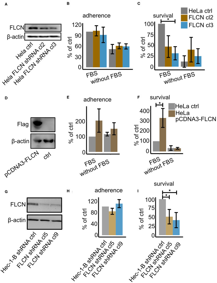
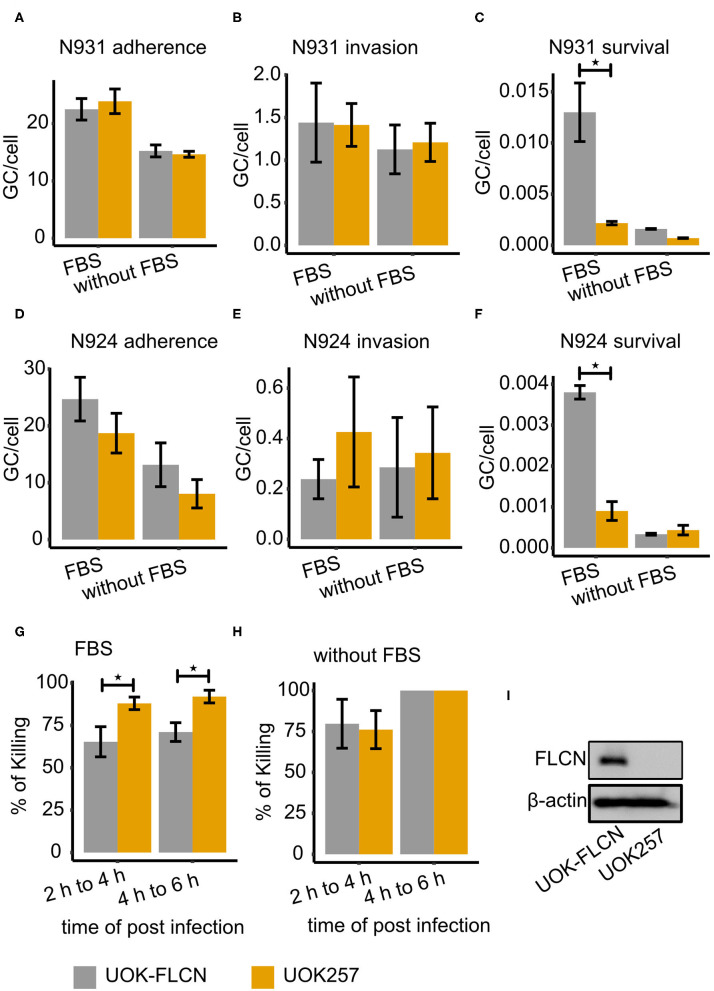
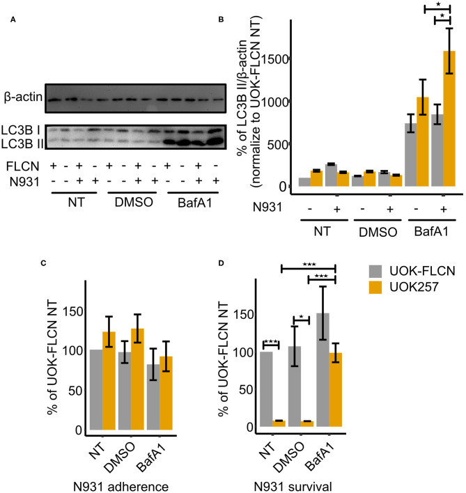
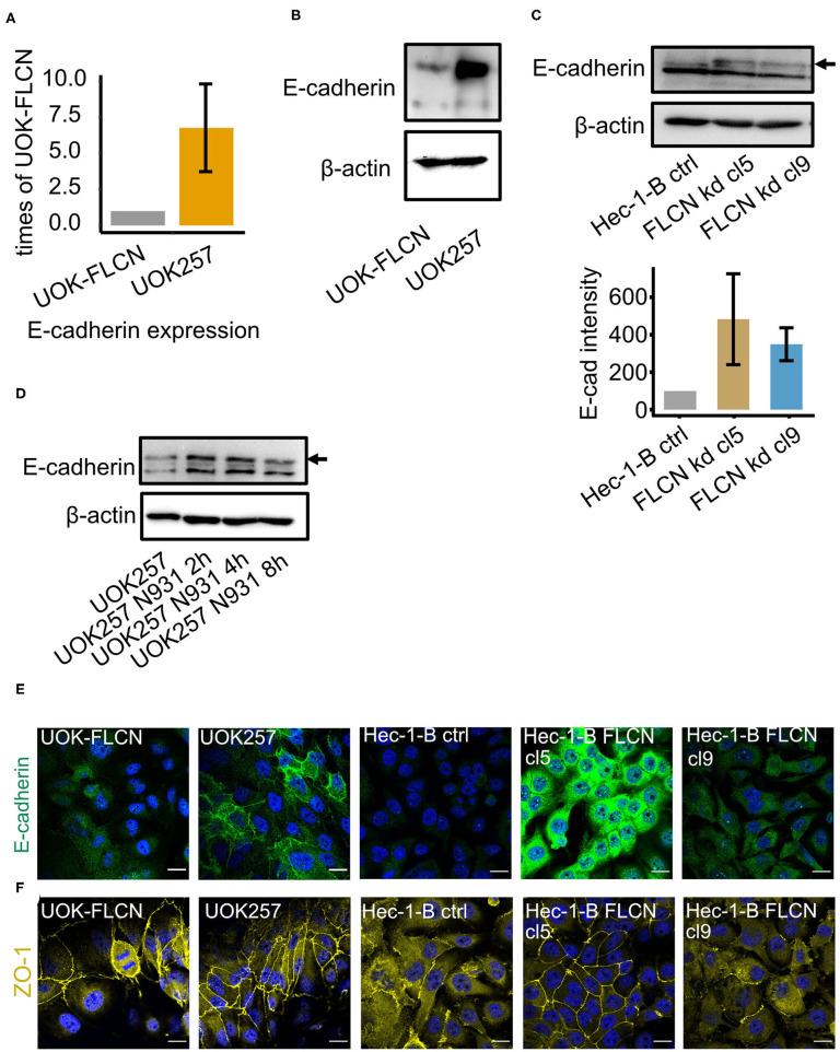
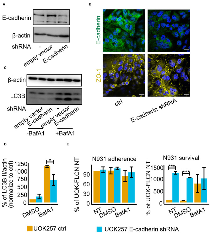
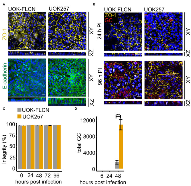
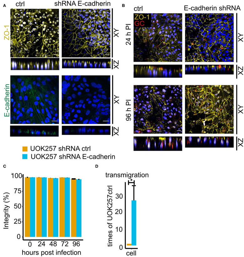
Neisseria gonorrhoeae, a Gram-negative obligate human pathogenic bacterium, infects human epithelial cells and causes sexually transmitted diseases. Emerging multi-antibiotic resistant gonococci and increasing numbers of infections complicate the treatment of infected patients. Here, we used an shRNA library screen and next-generation sequencing to identify factors involved in epithelial cell infection. Folliculin (FLCN), a 64 kDa protein with a tumor repressor function was identified as a novel host factor important for N. gonorrhoeae survival after uptake. We further determined that FLCN did not affect N. gonorrhoeae adherence and invasion but was essential for its survival in the cells by modulating autophagy. In addition, FLCN was also required to maintain cell to cell contacts in the epithelial layer. In an infection model with polarized cells, FLCN inhibited the polarized localization of E-cadherin and the transcytosis of gonococci across polarized epithelial cells. In conclusion, we demonstrate here the connection between FLCN and bacterial infection and in particular the role of FLCN in the intracellular survival and transcytosis of gonococci across polarized epithelial cell layers.

Keywords: autophagy; folliculin; gonococcal invasion; polarized cell culture; polarized epithelium.


Figures

Figure 1
Figure 1
FLCN downregulation leads to decreased survival of gonococci. HeLa2000 and Hec-1-B cells and their respective knockdown cell clones were infected with N931 (MOI 50) in RPMI or DMEM medium with or without FBS and then subjected to a gentamicin protection assay. (A) Western blot of FLCN of HeLa control and FLCN knockdown cells. (B) Infected cells were lysed and the total number of colonies forming units was quantified by dilution plating. Shown is the number of adhered bacteria (calculated as total minus surviving bacteria). (C) 50 μM gentamicin was added to the infected cells for 2 h to kill the extracellular gonococci. Cells were lysed and the number of colonies forming units was quantified. (D) Western blot of control and FLCN-Flag overexpressing HeLa2000 cells. Adhered (E) and intracellular (F) gonococci were determined and calculated by normalizing to HeLa control plus FBS. (G) Western blot of control and shRNA-expressing Hec-1-B cells. Adherent (H) and intracellular (I) bacteria were determined and normalized to control shRNA expressing Hec-1-B in DMEM plus FBS. Data (B,C,E,F,H,I) represent the mean ± s.d with three independent repeats. Significance was determined using student t-test, *P < 0.05.
Figure 2
Figure 2
FLCN supports gonococcal intracellular survival. Gonococcal adherence and survival were determined by gentamicin protection assay and invasion by differential immunostaining (see methods for details). Strains N931 (pili, porB1A-, Opa50+) and N924 (pili, porB1A-, Opa50) were used for infection. (A–C) Adherence, invasion and survival of N931 was determined in UOK-FLCN and UOK257 cells. (D–F) Adherence, invasion and survived of N924 in UOK-FLCN and UOK257 cells. (G,H) Shown are the rates by which intracellular N931 were killed in the respective cells grown in DMEM with (G) or without (H) FBS. Data represent the mean ± s.d with three independent repeats. Significance was determined using student t-test, *P < 0.05. (I) Western blot of UOK-FLCN and UOK257 to detect FLCN and actin was a loading control.
Figure 3
Figure 3
FLCN inhibits the autophagy flux and supports gonococcal intracellular survival. (A) Representative immunoblot displaying LC3B-I, LC3B-II, and β-actin of UOK-FLCN and UOK257 cells. The cells were either left untreated or were treated with the solvent DMSO or with 5 nM Bafilomycin A1 (BafA1) for 16 h in the absence or presence of gonococcal infection. (B) Densitometric quantification of LC3-II levels in three independent immunoblots shown in (A). Data represent the mean ± s.d with three independent repeats. Significance was determined using One-Way ANOVA, *P < 0.05. (C) Adherence and (D) intracellular survival of N931 were determined in UOK-FLCN and UOK257 either left untreated or treated with DMSO or 5 nM BafA1 for 16 h. The cells were infected with N931 (MOI 50) for 2 h and then analyzed by gentamicin protection assay. Data represent the mean ± s.d with three independent repeats. Significance was determined using student t-test, *P < 0.05, ***P < 0.001.
Figure 4
Figure 4
FLCN negatively regulates the E-cadherin in 2D cell culture. (A) qPCR of E-cadherin in UOK-FLCN and UOK257 cells. GAPDH was used as an internal control. (B) Western blot of UOK-FLCN and UOK257 cells to detect E-cadherin. Actin served as a loading control. (C) Western blot of the control and FLCN-knockdown Hec-1-B cells to detect E-cadherin and actin as a loading control. (D) UOK257 were infected with N931 for different time points and E-cadherin was analyzed by Western blotting. (E) Immunofluorescence staining of E-cadherin in UOK-FLCN, UOK257 and the control and FLCN knockdown cells of Hec-1-B. (F) Immunofluorescence staining of ZO-1 in UOK-FLCN, UOK257 and the control and FLCN knockdown cells of Hec-1-B.
Figure 5
Figure 5
E-cadherin knockdown in UOK257 cell increases gonococcal survival in 2D cell culture. (A) Western blot of E-cadherin in non-treated, empty vector-transfected and E-cadherin shRNA-transfected UOK257 cells. (B) Immunofluorescence staining of E-cadherin and ZO-1 in the control and E-cadherin knockdown cells of UOK257. (C) Representative immunoblot displaying LC3B-I, LC3B-II, and β-actin in the control and E-cadherin knockdown of UOK257 cells. The cells were treated with the solvent DMSO and 5 nM BafA1in DMSO for 16 h. (D) Densitometry quantification of LC3-II levels in three independent experiments [described in (C)]. LC3B-II in each lane was normalized to β-actin and the different experiment were normalized to the values obtained for the non-treated UOK257. Data represent the mean ± s.d with three independent repeats. Significance was determined using student t-test, *P < 0.05. (E) UOK257 were transfected with control or E-cadherin shRNAs and either treated with DMSO or 5 nM BafA1 in DMSO for 16 h. Then these cells were infected with gonococci for 2 h and adherence (left) and survival (right) was determined. Data represent the mean ± s.d with three independent repeats. Significance was determined using student t-test, ***P < 0.001.
Figure 6
Figure 6
FLCN negatively regulates E-cadherin and it delays gonococcal transmigration in polarized epithelial cells. (A) Confocal microscopy of transwell models of UOK-FLCN and UOK257. The cells were cultured for 10 days and then fixed and stained for E-cadherin or ZO-1 antibodies and DAPI. Z-stacks were made and reconstructed using FIJI. Shown were Z-projections (XY) and orthogonal sections (XZ). (B) Confocal microscopy of N931 infected (MOI 50) transwell models at different time points. Shown were Z-projections (XY) and orthogonal sections (XZ). (C) Barrier integrity of N931-infected (MOI 50) UOK-FLCN and UOK257 models were measured by FITC-dextran assay (4 kDa). Data represent the mean ± s.d with three independent repeats. (D) Transmigration of N931 (MOI 50) determined in UOK-FLCN and UOK257 transwell models. Data represent the mean ± s.d with three independent repeats, **P < 0.01.
Figure 7
Figure 7
Loss of E-cadherin enhances gonococcal transmigration in polarized epithelial cells. (A) Confocal microscopy of control and E-cadherin knockdown UOK257 cells grown transwells. The samples were stained for ZO-1 or E-cadherin and DAPI. Z-stacks were generated and reconstructed with FIJI. Shown are XY projections and orthogonal sections (XZ). (B) Confocal microscopy of N931-infected (MOI 50) transwell models at different time points. Shown are Z-projections (XY) and orthogonal sections (XZ). (C) Barrier integrity of N931-infected (MOI 50) UOK257 cells transfected with the vector control pLVTHM and E-cadherin shRNA in pLVTHM. Barrier integrity was determined by FITC-dextran assay (4 kDa). Data represent the mean ± s.d with three independent repeats. (D) Transmigration of N931 (MOI 50) determined in UOK257 transfected with pLVTHM and UOK257 E-cadherin shRNA after 48 h of infection. Data represent the mean ± s.d with three independent repeats, *P < 0.05.

Similar articles

Cited by

References

    1. Bos M. P., Grunert F., Belland R. J. (1997a). Differential recognition of members of the carcinoembryonic antigen family by Opa variants of Neisseria gonorrhoeae. Infect Immun 65:9. 10.1128/IAI.65.6.2353-2361.1997 - DOI - PMC - PubMed
    1. Bos M. P., Hogan D., Belland R. J. (1997b). Selection of Opa+ Neisseria gonorrhoeae by limited availability of normal human serum. Infect Immun 65:6. 10.1128/IAI.65.2.645-650.1997 - DOI - PMC - PubMed
    1. Chen T. (1995). Adherence of pilus− Opa+ gonococci to epithelial cells in vitro involves heparan sulfate. J. Exp. Med. 182, 511–517. 10.1084/jem.182.2.511 - DOI - PMC - PubMed
    1. Chen T., Gotschlich E. C. (1996). CGM1a antigen of neutrophils, a receptor of gonococcal opacity proteins. Proc. Natl. Acad. Sci. U.S.A. 93, 14851–14856. 10.1073/pnas.93.25.14851 - DOI - PMC - PubMed
    1. Criss A. K., Seifert H. S. (2006). Gonococci exit apically and basally from polarized epithelial cells and exhibit dynamic changes in type IV pili. Cell. Microbiol. 8, 1430–1443. 10.1111/j.1462-5822.2006.00722.x - DOI - PMC - PubMed

Publication types