

Dyspnea in Patient with Arteria Lusoria: A Case Report
- PMID: 33014911
- PMCID: PMC7515627
- DOI: 10.22038/ijorl.2020.46502.2525
Dyspnea in Patient with Arteria Lusoria: A Case Report
Abstract
Introduction: Arteria lusoria is an aberrant right subclavian artery. In symptomatic cases, patients report dysphagia and only in few cases dyspnea, due to external compression of the trachea and esophagus. Symptoms occur in advanced age and diagnosis is made with chest HRCT, when other causes of dysphagia have been excluded.
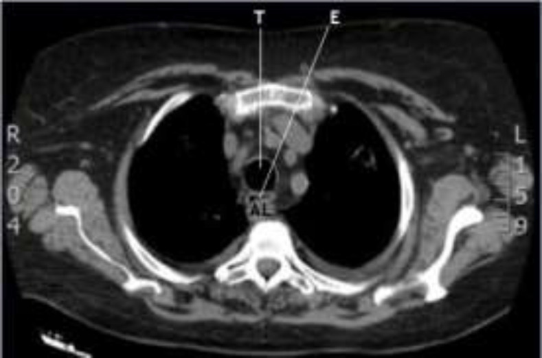
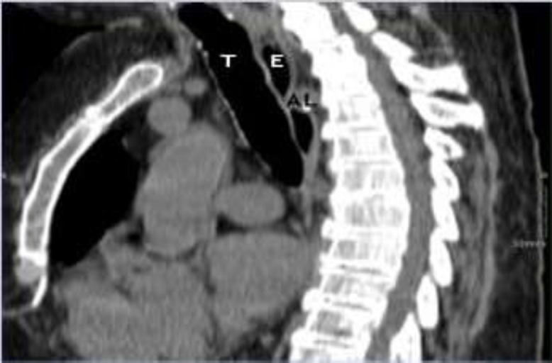
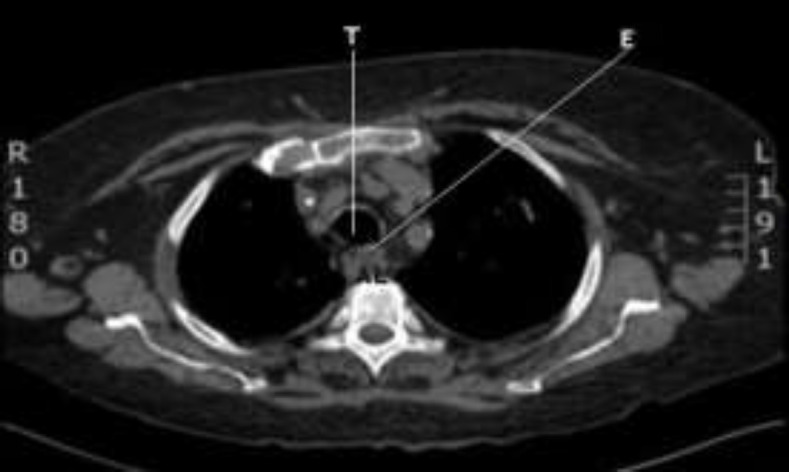
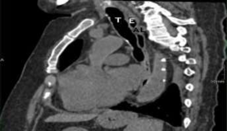
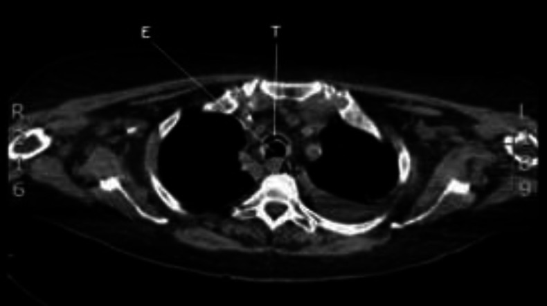
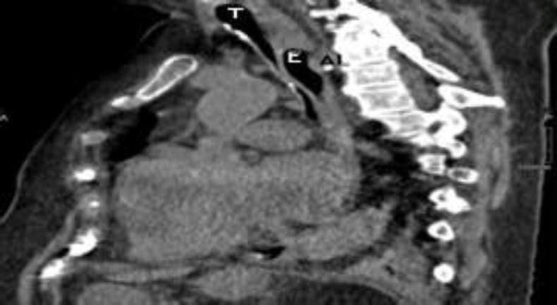
Case report: An 83-year-old woman presented with dyspnea and mechanical dysphagia for solids. Therefore, she did a chest high-resolution computed tomography (HRCT) that showed areas of consolidation of the lung parenchyma, pleural effusion and presence of arteria lusoria, with a retroesophageal course. After 18 days, dysphagia and dyspnea worsened. The new chest HRCT revealed bilateral atelectasis of the lower lung lobes and severe compression of esophagus and trachea along the course of the arteria lusoria.
Conclusion: Considering its dangerousness, this vascular anomaly should be considered in advanced aged patients with dysphagia and dyspnea, once other causes have been excluded.
Keywords: Arteria lusoria; Dysphagia; Dyspnea; Vascular anomaly.
Figures
Similar articles
-
Arteria Lusoria: An Unusual Cause of Dysphagia.Cureus. 2025 Mar 24;17(3):e81092. doi: 10.7759/cureus.81092. eCollection 2025 Mar. Cureus. 2025. PMID: 40271331 Free PMC article.
-
Dysphagia after arteria lusoria dextra surgery: Anatomical considerations before redo-surgery.World J Cardiol. 2017 Feb 26;9(2):191-195. doi: 10.4330/wjc.v9.i2.191. World J Cardiol. 2017. PMID: 28289534 Free PMC article.
-
[Arteria lusoria causing dyspnea: about a case].Pan Afr Med J. 2020 Dec 7;37:318. doi: 10.11604/pamj.2020.37.318.23253. eCollection 2020. Pan Afr Med J. 2020. PMID: 33654537 Free PMC article. French.
-
Arteria lusoria: developmental anatomy, clinical, radiological and surgical aspects.Ann Cardiol Angeiol (Paris). 2010 Jun;59(3):147-54. doi: 10.1016/j.ancard.2009.07.008. Epub 2009 Aug 8. Ann Cardiol Angeiol (Paris). 2010. PMID: 19962688 Review.
-
EUS imaging of the arteria lusoria: case series and review.Gastrointest Endosc. 2000 Nov;52(5):670-3. doi: 10.1067/mge.2000.109808. Gastrointest Endosc. 2000. PMID: 11060196 Review.
Cited by
-
Beyond the Classic Causes of Dysphagia: Bayford-Autenrieth Dysphagia.Cureus. 2024 Feb 23;16(2):e54755. doi: 10.7759/cureus.54755. eCollection 2024 Feb. Cureus. 2024. PMID: 38523923 Free PMC article.
References
-
- González-Sánchez M, Pardal-Refoyo J L, Martin-Sánchez A. The aberrant subclavian artery and disphagia lusoria. Acta Otorrinolaringol Esp. 2013;64:244–5. - PubMed
-
- Janssen M, Baggen MG, Veen HF, Smout AJ, Bekkers JA, Jonkman JG, et al. Dysphagia lusoria: clinical aspects, manometric findings, diagnosis, and therapy. Am J Gastroenterol. 2000;95:1411–16. - PubMed
-
- Derbel B, Saaidi A, Kasraoui R, Chaouch N, Aouini F. Aberrant right subclavian artery or arteria lusoria: a rare cause of dyspnea in children. Ann Vasc Surg. 2012;26(3):e1–4. - PubMed
-
- Atalaia-Martins C, Gonçalves C, Cotrim I, Pardal V. Dysphagia lusoria: a little-known cause of dysphagia. Rev Esp Enferm Dig. 2018;110(3):198–9. - PubMed
Publication types
LinkOut - more resources
Full Text Sources