. 2020 Sep 9:8:571004.
doi: 10.3389/fcell.2020.571004. eCollection 2020.

A Mutation in VWA1, Encoding von Willebrand Factor A Domain-Containing Protein 1, Is Associated With Hemifacial Microsomia

Affiliations

A Mutation in VWA1, Encoding von Willebrand Factor A Domain-Containing Protein 1, Is Associated With Hemifacial Microsomia

Yibei Wang et al. Front Cell Dev Biol. .

Abstract

Background: Hemifacial microsomia (HFM) is a type of rare congenital syndrome caused by developmental disorders of the first and second pharyngeal arches that occurs in one out of 5,600 live births. There are significant gaps in our knowledge of the pathogenic genes underlying this syndrome.

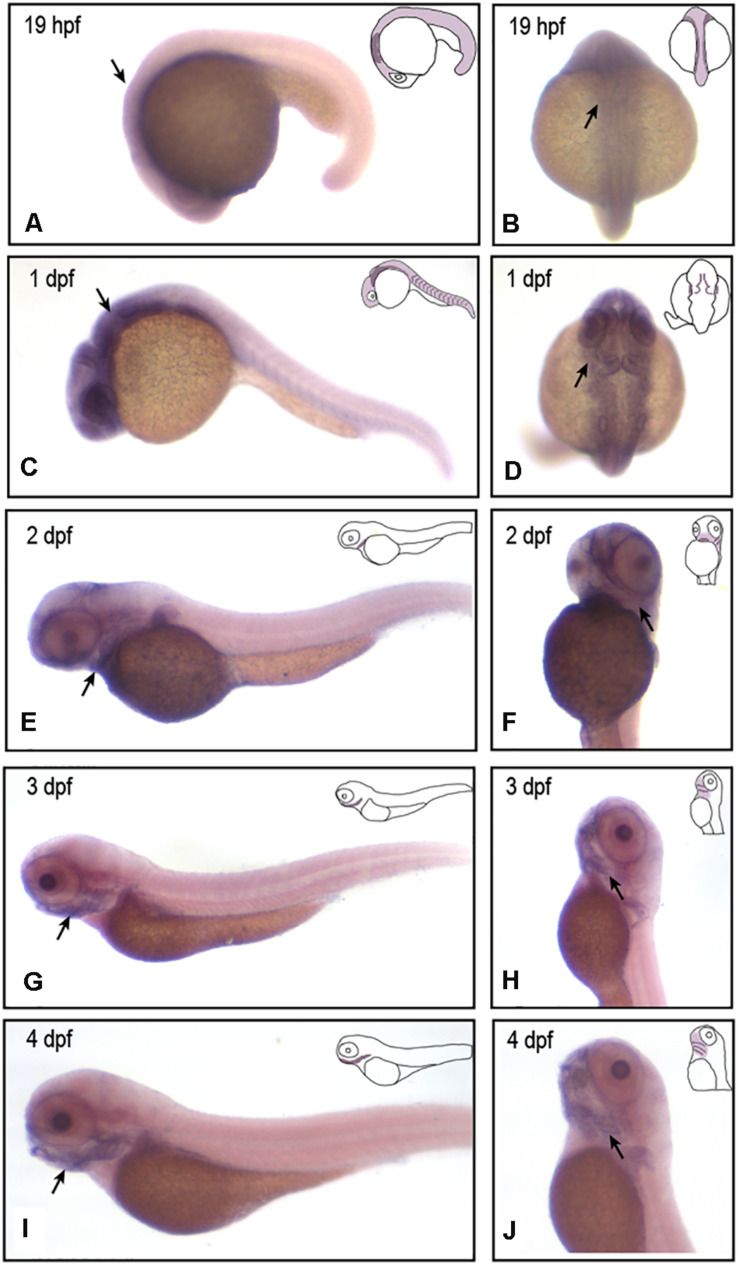
Methods: Whole exome sequencing (WES) was performed on five patients, one asymptomatic carrier, and two marry-in members of a five-generation pedigree. Structure of WARP (product of VWA1) was predicted using the Phyre2 web portal. In situ hybridization and vwa1-knockdown/knockout studies in zebrafish using morpholino and CRISPR/Cas9 techniques were performed. Cartilage staining and immunofluorescence were carried out.

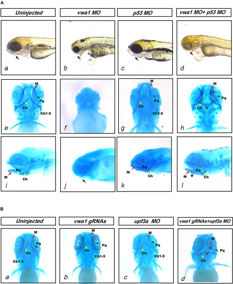
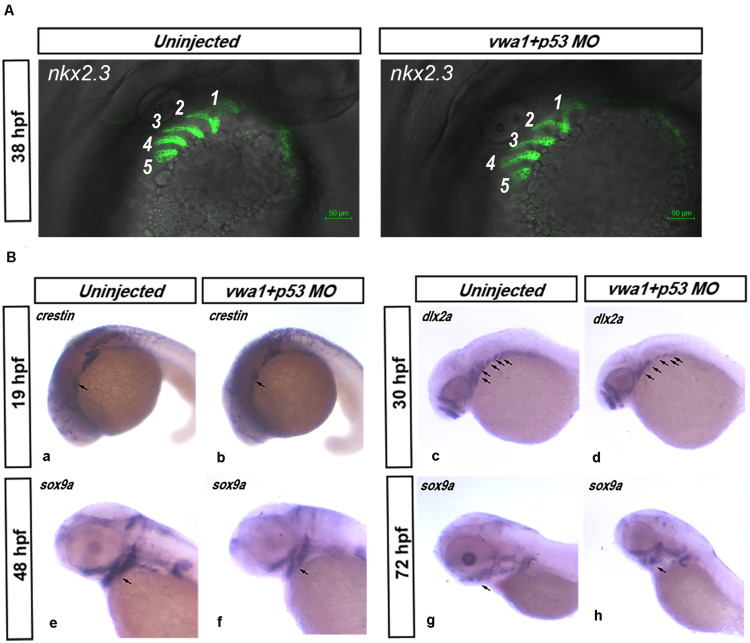
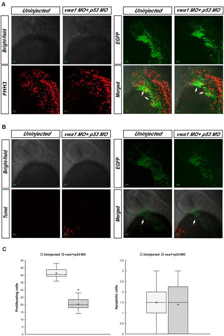
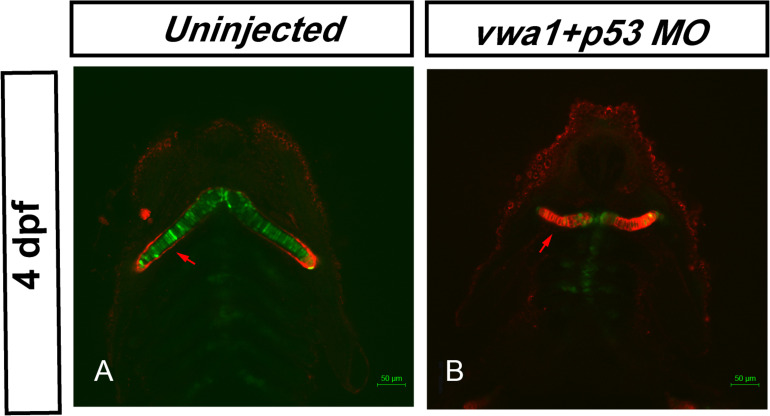
Results: Through WES and a set of filtration, we identified a c.G905A:p.R302Q point mutation in a novel candidate pathogenic gene, VWA1. The Phyre2 web portal predicted alterations in secondary and tertiary structures of WARP, indicating changes in its function as well. Predictions of protein-to-protein interactions in five pathways related to craniofacial development revealed possible interactions with four proteins in the FGF pathway. Knockdown/knockout studies of the zebrafish revealed deformities of pharyngeal cartilage. A decrease of the proliferation of cranial neural crest cells (CNCCs) and alteration of the structure of pharyngeal chondrocytes were observed in the morphants as well.

Conclusion: Our data suggest that a mutation in VWA1 is functionally linked to HFM through suppression of CNCC proliferation and disruption of the organization of pharyngeal chondrocytes.

Keywords: VWA1; cranial neural crest cell; hemifacial microsomia; whole exome sequencing; zebrafish.


Figures

FIGURE 1
FIGURE 1
HFM pedigree and the identification of vwa1. (A) Diagram of the five-generation HFM pedigree. This is a five-generation Chinese kinship in which 8 of 11 direct relatives displayed HFM to varying degrees. The proband showed grade III bilateral ear deformities; individual III:1 in the pedigree had a grade II unilateral ear deformity, whereas other patients exhibited unilateral grade III ear deformities. All patients exhibited mandibular hypoplasia. Circles denote females and squares denote males. The text immediately below the symbols shows the ID of the corresponding family member; formula image 
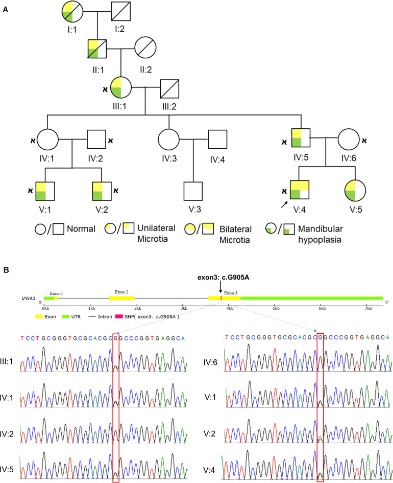
indicates that whole-exome sequencing was done. (B) Sanger sequencing results of the VWA1 mutation in the pedigree. Patients III:1, IV:5, V:1, V:2 and V:4, and the asymptomatic carrier IV:1, all had the same non-synonymous point mutation (c.G905A:p.R302Q) in VWA1, whereas the two marry-in members of the family (IV:2 and IV:6) did not.
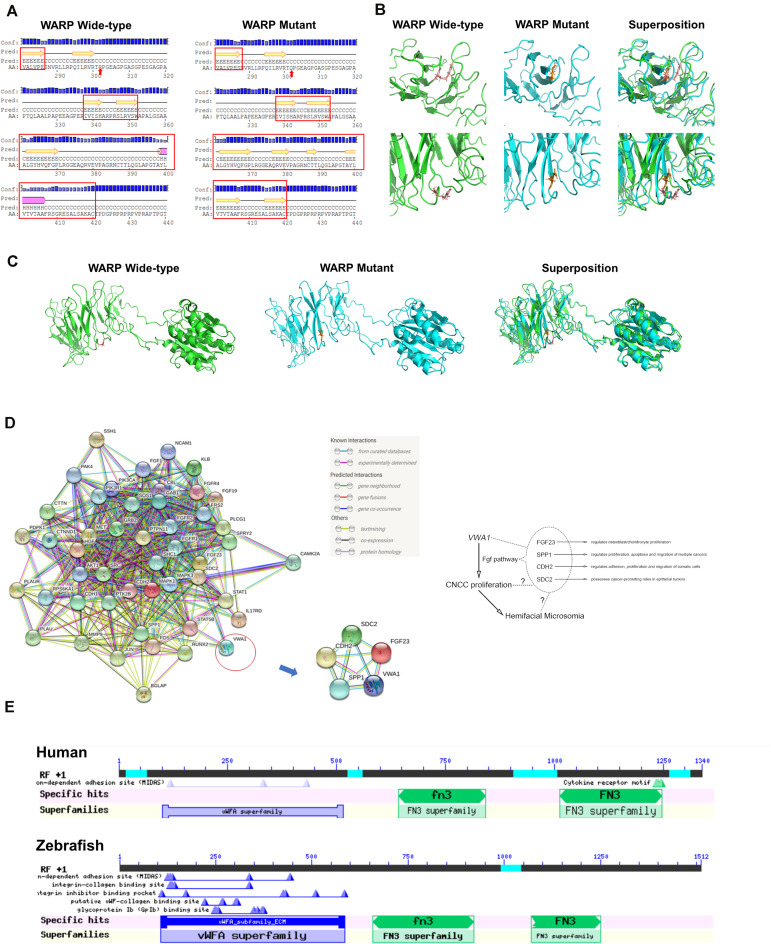
FIGURE 2
FIGURE 2
Predicted WARP structures, protein interactions and conserved domains. (A) Predicted secondary structures of wild-type and mutant protein sequences flanking the mutation. Secondary structural features are annotated as follows: pink cylinder, α-helix; yellow arrow, β-sheet; black line, coil; Conf, confidence; Pred, predicted; H in Pred line, helix; C in Pred line, coil; E in Pred line, sheet; AA, amino acid; red arrow, mutant amino acid. Local (B) and global (C) views of the predicted tertiary structures of wild-type (green) and mutant (blue) proteins. Structural superposition analyses demonstrating the change in protein structure brought about by the mutation are also displayed. (D) Protein-to-protein interactions of WARP with the FGF pathway. Predictions using the web-based program, String, indicated that WARP may functionally interact with four proteins of FGF pathway: FGF23, SPP1, CDH2, and SDC2. (E) Domains of WARP conserved in humans and zebrafish. Humans and zebrafish have the same WARP conserved domains: a von Willebrand factor A-domain, the first fibronectin type III repeat, and the second fibronectin type III repeat.
FIGURE 3
FIGURE 3
vwa1 expression in developing zebrafish embryos. In situ hybridization (ISH) results showing the expression of vwa1 at 19 hpf (A,B), 1 dpf (C,D), 2 dpf (E,F), 3 dpf (G,H) and 4 dpf (I,J) stages in lateral (A,C,E,G,I), dorsal (B,D) and lateral-ventral (F,H,J) views. At 19 hpf and 1 dpf, vwa1 was expressed throughout the body, with expression in the head being higher than that in other parts of the body (black arrows in A,C). At 19 hpf and 1 dpf, vwa1 expression was detected at the medial edge of the pharyngeal arch (black arrows in B,D). At 2 dpf, vwa1 staining in the head began to increase slightly, especially in the mandible (black arrow in E). At 3 dpf and 4 dpf, expression of vwa1 was restricted to the mandible (black arrows in G,I). Staining of vwa1 in the pharyngeal pouch was stronger than that in the pharyngeal arch (black arrows in F–H). vwa1 was also expressed in the epidermis of the brain (D) and in somites (C,E,G,I).
FIGURE 4
FIGURE 4
Deformities of pharyngeal cartilage in vwa1 morphants and mutants. (A) Deformities of pharyngeal cartilage in vwa1 morphants. The figure shows the jaws of uninjected and vwa1 morpholino (MO)-, p53 MO- and vwa1 + p53 MO-injected morphants under a stereoscope and Alcian blue staining of morphant embryos. Injection of 1 ng vwa1-ATG MO caused jaw structures to almost completely disappear (b, f, and g). Injection of 1 ng vwa1-ATG MO with p53 MO reduced the overall size of jaw structures at 4 dpf compared with uninjected controls and 1.5 ng p53 MO-injected morphants (a and d), whereas there was no significant difference compared with controls after injection of p53 MO (a–d and i–l). The first and second pharyngeal arches were severely hypoplastic (brown ⋆ in h and l), and the third to seventh pharyngeal arches (Cb1-5, circled in e) were absent (h). Joint junctions were abnormal: the angle between cartilage derived from the first pharyngeal arch, Meckel’s (M) and palatoquadrate (Pq) became smaller, whereas the angle between cartilage derived from the second pharyngeal arch ceratohyal (Ch) became larger (brown ⋆ in h and l). (B) Deformities of pharyngeal cartilage in vwa1-knockout mutants. a, b, c, and d shows Alcian blue staining results for uninjected embryos and embryos injected with vwa1 gRNAs, upf3a MO, and vwa1 gRNAs together with upf3a MO. Embryos injected with vwa1 gRNAs or upf3a MO showed no significant deformities in pharyngeal cartilage compared with uninjected embryos (b and c). Injection of vwa1 gRNAs together with upf3a MO resulted in pharyngeal cartilage deformities similar to those in vwa1 morphants, including hypoplasia of first and second pharyngeal cartilages, disappearance of the third to seventh pharyngeal arches, and abnormal joint junctions (brown ⋆ in d).
FIGURE 5
FIGURE 5
Effects of reduced vwa1 on the development of pharyngeal cartilage. (A) Endodermal pouch in Tg (nkx2.3:EGFP) embryos. At about 38 hpf, when the endodermal pouch is almost fully formed, no apparent difference was observed in the pharyngeal pouch in vwa1 morphants (right) compared with uninjected morphants (left). (B) vwa1 knockdown causes a reduction in cranial neural crest cell numbers. At about 19 hpf, when neural crest cells migrate to the cranial area, the expression of crestin in vwa1-knockdown embryos was not obviously different from that of uninjected control embryos (a and b). At 30 hpf, when migration of neural crest cells is complete, dlx2a was expressed in similar domains, but the area of these domains was reduced significantly in vwa1 morphants (c and d). At 48 and 72 hpf, when the cranial neural crest has differentiated into pharyngeal chondrocytes, sox9a was expressed in similar domains, but the areas of these domains were reduced significantly in vwa1 morphants (e–h). These data show that, in vwa1 morphants, neural crest cells generally could migrate to the pharyngeal arch and specify to chondrocytes, but their numbers were reduced.
FIGURE 6
FIGURE 6
Proliferation and apoptosis of cranial neural crest cells at approximately 30 hpf. (A) Immunofluorescence results showing anti-phosphohistone H3 (PHH3) staining demonstrating cell proliferation. Merged image demonstrates higher anti-PHH3 signals in cranial neural crest cells compared with uninjected embryos. Arrows: pharyngeal arches. (B) Apoptosis assessed by immunofluorescence detection of TUNEL assay. The apoptosis signal in the vwa1 + p53 MO group did not coincide with EGFP fluorescence. Arrows: pharyngeal arches. (C) Comparison of apoptotic and proliferative numbers of neural crest cells in pharyngeal arches between vwa1 + p53 MO-injected embryos and uninjected control embryos. The difference in cell proliferation was statistically significant (p < 0.05, Student’s t-test). Lower/upper whiskers: 1st and 4th quartiles of the data; box: 2nd and 3rd quartiles of the data; mid-line: median of the data; ×: mean value of the data; *: significantly different.
FIGURE 7
FIGURE 7
Knockdown of vwa1 leads to disorganization of pharyngeal chondrocytes at 4 dpf. (A) In uninjected control embryos at 4 dpf, craniofacial cartilage had formed and showed a very stereotypic shape between individuals, exhibiting the characteristic “stack of pennies” organization in which thin and elongated chondrocytes are assembled on top of one another to form their respective cartilage element. (B) In vwa1 morphants, the size and length-width ratio of many cartilage chondrocytes were smaller and the cartilage elements were greatly deformed compared with those of uninjected controls.

Similar articles

Cited by

References

    1. Akimenko M. A., Ekker M., Wegner J., Lin W., Westerfield M. (1994). Combinatorial expression of three zebrafish genes related to distal-less: part of a homeobox gene code for the head. J. Neurosci. 14 3475–3486. 10.1523/jneurosci.14-06-03475.1994 - DOI - PMC - PubMed
    1. Allen J. M., Bateman J. F., Hansen U., Wilson R., Bruckner P., Owens R. T., et al. (2006). WARP is a novel multimeric component of the chondrocyte pericellular matrix that interacts with perlecan. J. Biol. Chem. 281 7341–7349. 10.1074/jbc.m513746200 - DOI - PubMed
    1. Allen J. M., Brachvogel B., Farlie P. G., Fitzgerald J., Bateman J. F. (2008). The extracellular matrix protein WARP is a novel component of a distinct subset of basement membranes. Matrix Biol. 27 295–305. 10.1016/j.matbio.2008.01.005 - DOI - PubMed
    1. Allen J. M., Zamurs L., Brachvogel B., Schlötzer-Schrehardt U., Hansen U., Lamandé S. R., et al. (2009). Mice lacking the extracellular matrix protein WARP develop normally but have compromised peripheral nerve structure and function. J. Biol. Chem. 284 12020–12030. 10.1074/jbc.M806968200 - DOI - PMC - PubMed
    1. Babb-Clendenon S., Shen Y.-C., Liu Q., Turner K. E., Mills M. S., Cook G. W., et al. (2006). Cadherin-2 participates in the morphogenesis of the zebrafish inner ear. J. Cell Sci. 119(Pt 24), 5169–5177. 10.1242/jcs.03299 - DOI - PubMed

LinkOut - more resources