. 2020 Jul 20;7(10):270-285.
doi: 10.15698/mic2020.10.732.

Variants of the human RAD52 gene confer defects in ionizing radiation resistance and homologous recombination repair in budding yeast

Affiliations

Variants of the human RAD52 gene confer defects in ionizing radiation resistance and homologous recombination repair in budding yeast

Alissa D Clear et al. Microb Cell. .

Abstract

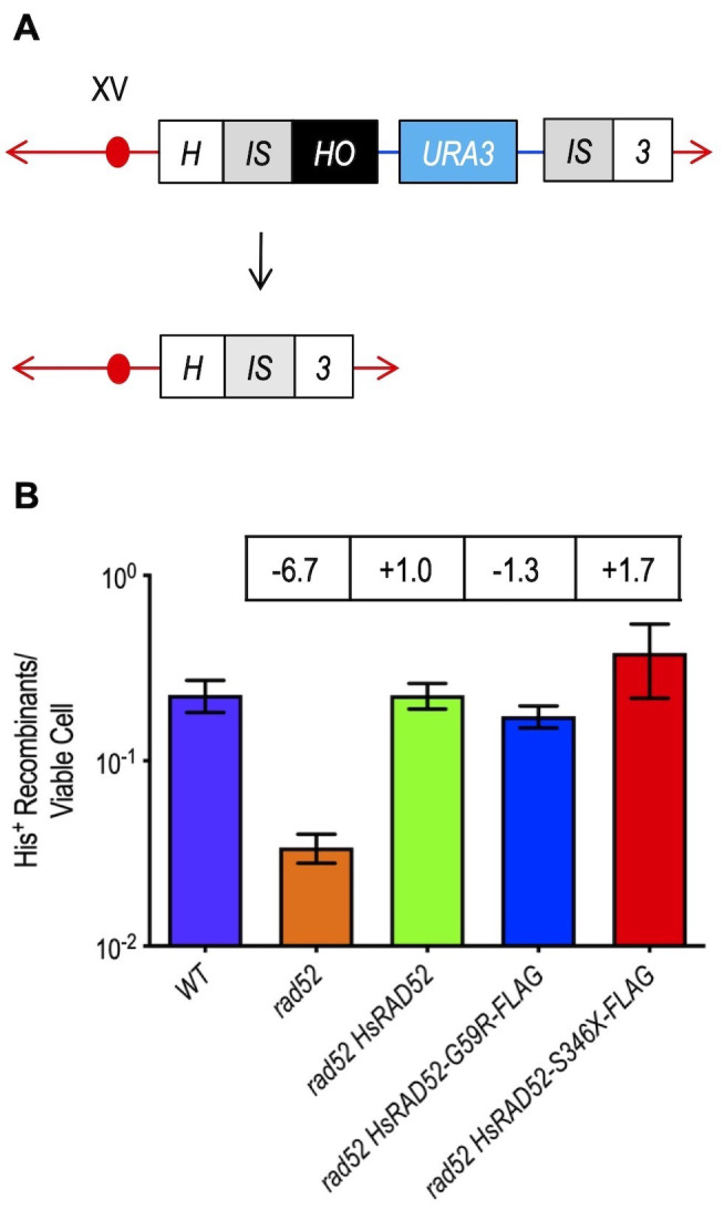
RAD52 is a structurally and functionally conserved component of the DNA double-strand break (DSB) repair apparatus from budding yeast to humans. We recently showed that expressing the human gene, HsRAD52 in rad52 mutant budding yeast cells can suppress both their ionizing radiation (IR) sensitivity and homologous recombination repair (HRR) defects. Intriguingly, we observed that HsRAD52 supports DSB repair by a mechanism of HRR that conserves genome structure and is independent of the canonical HR machinery. In this study we report that naturally occurring variants of HsRAD52, one of which suppresses the pathogenicity of BRCA2 mutations, were unable to suppress the IR sensitivity and HRR defects of rad52 mutant yeast cells, but fully suppressed a defect in DSB repair by single-strand annealing (SSA). This failure to suppress both IR sensitivity and the HRR defect correlated with an inability of HsRAD52 protein to associate with and drive an interaction between genomic sequences during DSB repair by HRR. These results suggest that HsRAD52 supports multiple, distinct DSB repair apparatuses in budding yeast cells and help further define its mechanism of action in HRR. They also imply that disruption of HsRAD52-dependent HRR in BRCA2-defective human cells may contribute to protection against tumorigenesis and provide a target for killing BRCA2-defective cancers.

Keywords: DNA double strand breaks; HsRAD52 variants; budding yeast; homologous recombination repair; ionizing radiation; tumorigenesis.


Conflict of interest statement

Conflict of interest: The authors declare that there are no conflicts of interest.

Figures

Figure 1
Figure 1. FIGURE 1: The adh1::HsRAD52-G59R-FLAG and adh1::HsRAD52-S346X-FLAG alleles express stable proteins in budding yeast cells.
Whole cell extracts from strains ABX3684-12B (adh1::HsRAD52-FLAG), ABX3782-2D (adh1::HsRAD52-G59R-FLAG) and ABX3974-11C (adh1::HsRAD52-S346X-FLAG) were run on SDS-PAGE gels, blotted to a nylon membrane and probed with anti-FLAG and anti-GAPDH antibodies. The genotypes of the strains are denoted at the top of the figure. Bands corresponding to wild-type and mutant HsRAD52, and GAPDH are labeled on the left side of the figure.
Figure 2
Figure 2. FIGURE 2: The adh1::HsRAD52-G59R-FLAG and adh1::HsRAD52-S346X-FLAG alleles fail to suppress the ionizing radiation sensitivity of rad52-/- mutant yeast cells.
Cultures of wild-type (ABX3566), rad52-/- (ABX3568), rad52-/- adh1::HsRAD52-FLAG+/+ (ABX4130), rad52-/- adh1::HsRAD52-G59R-FLAG-/- (ABX4129) and rad52-/- adh1::HsRAD52-S346X-FLAG-/- (ABX4131) yeast strains grown to mid-log phase were counted before being subjected to 20, 40, 80, 160 and 320 Gy of X-ray radiation. Appropriate dilutions of unirradiated and irradiated cultures were plated onto solid YPD medium, incubated at 30°C for three days, and the resulting colonies counted. Percent viability was calculated by dividing the number of colonies arising on the plates by the number of cell bodies plated and multiplying by 100. Mean percent survival was calculated for at least 10 independent cultures for each genotype. These values and 95% confidence intervals were plotted against levels of radiation exposure.
Figure 3
Figure 3. FIGURE 3: The adh1::HsRAD52-G59R-FLAG and adh1::HsRAD52-S346X-FLAG alleles do not suppress the MTI defect of rad52 mutant yeast cells.
(A) Cartoon depicting MTI. DSB formation by HO endonuclease cutting at the HO cut site on the centromere distal edge of the Ya sequence (blue box) at the MAT locus on chromosome III precipitates the exonucleolytic removal of “a” mating type information prior to its replacement by unidirectional transfer of “α” mating type information from the Yα sequence (red box) of the flanking, intact but silent HML locus. This results in the switching of the cell from the “a” mating type to the “α” mating type. (B) The adh1::HsRAD52-G59R-FLAG and adh1::HsRAD52-S346X-FLAG alleles confer defects in MTI. Single colonies of haploid wild-type (ABX3817-15B), rad52 (ABX3817-7D), rad52 adh1::HsRAD52-FLAG (ABX3977-10C), rad52 adh1::HsRAD52-G59R-FLAG (ABX3985-55B), and rad52 adh1::HsRAD52-S346X (ABX3994-18D) strains were used to inoculate at least 10 one milliliter YPGL cultures and grown overnight. Following a period of expression of HO endonuclease, appropriate dilutions were plated onto YPD medium, incubated for three days at 30°, and the number of colonies counted. Colonies were replica plated to fresh YPD plates, printed with a lawn of the haploid R113a mating type tester strain, printed plates incubated overnight at 30°C and then replica plated onto SD plates, which were incubated overnight at 30°C. Frequencies of MTI were determined by dividing the number of diploid patches arising on the SD plates by the number of colonies counted on the original YPD plates. Mean frequencies of MTI and 95% confidence intervals were plotted against genotype. Fold differences below (-) the wild-type frequency of MTI for each strain are indicated in boxes above the bar for each mean frequency.
Figure 4
Figure 4. FIGURE 4: The adh1::HsRAD52-G59R-FLAG and adh1::HsRAD52-S346X-FLAG alleles complement the loss of DSB repair by SSA in rad52 mutant yeast cells.
(A) Cartoon depicting DSB repair by recombination between non-tandem direct repeats. At the HIS3 locus on chromosome XV, DSB formation by HO endonuclease cutting at a HO cut site (black box) inserted at the right edge of the left duplication of a segment of the HIS3 coding sequence (left gray IS box) initiates bidirectional exonucleolytic processing. Processing reveals complementary single-stranded sequences at the left and right repeats (left and right gray IS boxes) that anneal, creating non-homologous tails whose removal deletes intervening plasmid sequences (blue line and aqua URA3 marker box) enroute to creating an intact HIS3 gene. (B) The adh1::HsRAD52-G59R-FLAG and adh1::HsRAD52-S346X-FLAG alleles complement the defects in DSB repair by DRR. Single colonies of haploid wild-type (ABM325), rad52 (ABM326), rad52 adh1::HsRAD52 (ABM507), rad52 adh1::HsRAD52-G59R-FLAG (ABX3970-88A), and rad52 adh1::HsRAD52-S346X (ABX3975-15A) strains were used to inoculate at least 10 one milliliter YPGL cultures and grown overnight. After a period of expression of HO endonuclease, appropriate dilutions were plated onto solid YPD medium to determine viability, and onto medium lacking histidine to select for recombinants. Following incubation for three days at 30° colonies were counted and frequencies of DRR determined by dividing the number of His+ recombinants by the number of viable cells plated. Mean frequencies of DRR and 95% confidence intervals were plotted against genotype. Fold differences above (+) and below (-) wild-type are indicated in the boxes above the bar for each mean frequency.
Figure 5
Figure 5. FIGURE 5: HsRAD52-S346X alters the in-solution oligomerization state of HsRAD52.
Superdex 200 size exclusion elution profiles are shown for HsRAD521-212, HsRAD52-G59R1-212, and HsRAD52-S346X. HsRAD521-212 (red trace) eluted as a single peak at 50.0ml, with an estimated size of 360kDa. HsRAD52-G59R1-212 (green trace) eluted as a single peak at 49.6 ml with an estimated size of 366kDa. HsRAD52-S346X (blue trace) eluted as two peaks; the first peak eluted at 51.9 ml with an estimated size of 335kDa, and the second peak eluted at 58.7ml with an estimated size of 257kDa. (mAU = milliabsorbance unit).
Figure 6
Figure 6. FIGURE 6: The adh1::HsRAD52-G59R-FLAG and adh1::HsRAD52-S346X-FLAG alleles have differential effects on the interaction of HsRAD52 with the MAT and HML loci during MTI.
(A) Cartoon depicting substrates for MTI and location of primers used for quantitation of immunoprecipitated genomic sequences. Following DSB formation by HO endonuclease at the HO cut site (black box) at the MAT locus on chromosome III, exonucleolytic processing results in the accumulation of ssDNA in flanking sequences. ScRad52-FLAG, or HsRAD52-FLAG progressively associate with the ssDNA. This association facilitates retention of the DNA sequences by ChIP, which are quantitated by qPCR using MATa recipient primers (red arrows; Table S3) complementary to a 110 bp sequence laying 500 bp upstream from the DSB at MAT. Deletion of the HMR locus and replacement with a hygMX marker (purple box) removes additional complementary sequences from the genome. After association with ssDNA at MAT, the HRR apparatus facilitates a search for homologous genomic sequences that gives rise to heteroduplex formation with the intact HML locus. The sequences that lay proximal to the HO cut site at the border of Yα (red box) are the putative initial location for heteroduplex formation. Association of ScRad52-FLAG or HsRAD52-FLAG with the heteroduplex results in retention of these sequences by ChIP, and their quantitation is done by qPCR using HMLα donor primers (red arrows; Table S3) complementary to a 187 bp sequence laying 67 bp downstream from the HO cut site sequence at HML. (B) ScRad52-FLAG and HsRAD52-FLAG display similar kinetics of association with sequences at the MAT locus after DSB formation. Single colonies of wild-type (ABX3961-4C), rad52 (ABX3943-3B), rad52 adh1::HsRAD52-FLAG (ABX3977-10C), rad52 adh1::HsRAD52-G59R-FLAG (ABX3985-55B), and rad52 adh1::HsRAD52-S346X-FLAG (ABX3994-18D) strains carrying MTI assay components were used to establish cultures from which aliquots were collected at various times before and after DSB formation at the MAT locus by HO endonuclease. Whole cell extracts were prepared, subjected to ChIP using anti-FLAG antibody and the immunoprecipitated DNA from the MAT (experimental) and SAM1 (control) loci quantitated by qPCR. Fold changes in degree of occupancy of the FLAG-tagged proteins relative to those observed before DSB formation were normalized to a control strain lacking FLAG-tagged proteins (ABX3933-46C). Mean fold changes from at least eight determinations using DNA collected from at least three independent time courses, and standard deviations were plotted against elapsed time after initiation of DSB formation. (C) HsRAD52-G59R-FLAG and HsRAD52-S346X-FLAG display defects in association with the HMR locus during MTI. Same as above except immunoprecipitated DNA from the HMR locus (experimental) was quantitated by qPCR.
Figure 7
Figure 7. FIGURE 7: The adh1::HsRAD52-G59R-FLAG and adh1::HsRAD52-S346X-FLAG alleles confer defects in repair synthesis during MTI.
(A) Cartoon depicting extension by repair DNA synthesis of putative heteroduplex formed upon association between sequences at the MAT and HML loci during MTI. Following DSB formation at the MAT locus (blue lines) exonucleolytic processing creates ssDNA onto which ScRad52-FLAG and HsRAD52-FLAG are proposed to bind. A search for homology putatively results in formation of a heteroduplex linking homologous sequences at the broken MAT locus with sequences at the intact HML locus (solid red lines). Extension of the heteroduplex by DNA repair synthesis (dotted red line) copies “α” information from HML that replaces “a” information lost from MAT, ultimately resulting in a change from MATa to MATα. Repair synthesis ultimately covalently joins “α” information from HML with sequences downstream from the MAT locus, which can be detected by PCR using the depicted primers (black arrows). (B) Repair synthesis was defective in rad52 adh1::HsRAD52-G59R-FLAG and rad52 adh1::HsRAD52-S346X-FLAG mutant cells. Genomic DNA was collected before immunoprecipitation from the same wild-type (ABX3961-4C), rad52 adh1::HsRAD52-FLAG (ABX3977-10C), rad52 adh1::HsRAD52-G59R-FLAG (ABX3985-55B), and rad52 adh1::HsRAD52-S346X-FLAG (ABX3994-18D) cultures used for the ChIP analyses described above (Fig 6). Extension by repair synthesis of the putative MAT/HML heteroduplex was quantitated by semi-quantitative end-point PCR using the primers depicted in panel A. DNA from the intact SAM1 locus was also quantitated as a signal for normalization. PCR products were separated on agarose gels, stained with ethidium bromide and band intensities were quantified using ImageJ. Normalized mean ratios and corresponding standard deviations from three independent time courses were calculated by dividing signal obtained from repair synthesis with the signal from the SAM1 control. All time point values were then normalized to the signal obtained before DSB formation (T=0 hrs) and plotted against elapsed time after DSB formation.

Similar articles

Cited by

References

    1. Frankenberg-Schwager M, Frankenberg D. DNA double-strand breaks: their repair and relationship to cell killing in yeast. Int J Radiat Biol. 1990;58(4):569–575. doi: 10.1080/09553009014551931. - DOI - PubMed
    1. Khanna KK, Jackson SP. DNA double-strand breaks: signaling, repair and the cancer connection. Nat Genet. 2001;27(3):247–254. doi: 10.1038/85798. - DOI - PubMed
    1. Pastink A, Eeken JC, Lohman PH. Genomic integrity and the repair of double-strand DNA breaks. Mutat Res. 2001;480-481:37–50. doi: 10.1016/s0027-5107(01)00167-1. - DOI - PubMed
    1. Santivasi WL, Xia F. Ionizing radiation-induced DNA damage, response, and repair. Antioxid Redox Signal. 2014;21(2):251–259. doi: 10.1089/ars.2013.5668. - DOI - PubMed
    1. Essers J, Hendriks RW, Swagemakers SM, Troelstra C, de Wit J, Bootsma D, Hoeijmakers JH, Kanaar R. Disruption of mouse RAD54 reduces ionizing radiation resistance and homologous recombination. Cell. 1997;89(2):195–204. doi: 10.1016/s0092-8674(00)80199-3. - DOI - PubMed

LinkOut - more resources