. 2020 Sep 16:2020:3634974.
doi: 10.1155/2020/3634974. eCollection 2020.

Protective Role of Tangshen Formula on the Progression of Renal Damage in db/db Mice by TRPC6/Talin1 Pathway in Podocytes

Affiliations

Protective Role of Tangshen Formula on the Progression of Renal Damage in db/db Mice by TRPC6/Talin1 Pathway in Podocytes

Qian Wang et al. J Diabetes Res. .

Abstract

Tangshen Formula (TSF) is a Chinese Medicine formula that has been reported to alleviate proteinuria and protect renal function in humans and animals with diabetic kidney disease (DKD). However, little is known about its mechanism in improving proteinuria. The dysregulation of podocyte cell-matrix adhesion has been demonstrated to play an important role in the pathogenesis and progression of proteinuric kidney diseases including DKD. In the present study, the underlying protective mechanism of TSF on podocytes was investigated using the murine model of type 2 DKD db/db mice in vivo and advanced glycation end products (AGEs)-stimulated primary mice podocytes in vitro. Results revealed that TSF treatment could significantly mitigate reduction of podocyte numbers and foot process effacement, reduce proteinuria, and protect renal function in db/db mice. There was a significant increase in expression of transient receptor potential canonical channel 6 (TRPC6) and a decrease in expression of talin1 in podocytes of db/db mice. The results of AGEs-stimulated primary mice podocytes showed increased cell migration and actin-cytoskeleton rearrangement. Moreover, primary mice podocytes stimulated by AGEs displayed an increase in TRPC6-dependent Ca2+ influx, a loss of talin1, and translocation of nuclear factor of activated T cell (NFATC) 2. These dysregulations in mice primary podocytes stimulated by AGEs could be significantly attenuated after TSF treatment. 1-Oleoyl-2-acetyl-sn-glycerol (OAG), a TRPC6 agonist, blocked the protective role of TSF on podocyte cell-matrix adherence. In conclusion, TSF could protect podocytes from injury and reduce proteinuria in DKD, which may be mediated by the regulation of the TRPC6/Talin1 pathway in podocytes.


Conflict of interest statement

The authors have no conflicts of interest to declare.

Figures
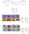
Figure 1
Figure 1
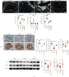
TSF reduced proteinuria and alleviated renal injury in db/db mice. (a) Body weight was recorded each week (n = 6). (b) Kidney weight of mice in each group at 22 weeks of age (n = 6). (c) Urinary albumin to creatinine ratio (ACR) (n = 6). (d) Serum creatinine in each group (n = 6). (e) Representative PAS staining (scale bars, 20 μm) and quantitative analysis of glomerular hypertrophy and mesangial expansion (n = 6); representative PASM staining (scale bars, 20 μm). (f) Representative immunohistochemical staining and quantitative analysis of collagen type IV and fibronectin (scale bars, 20 μm) (20 glomeruli were randomly evaluated per section) (n = 6). The data were expressed as the mean ± SEM. #P < 0.05, ##P < 0.01 vs. db/m group; P < 0.05, ∗∗P < 0.01 vs. db/db group. Statistically analyzed via a one-way ANOVA with Dunnett's correction.
Figure 2
Figure 2
TSF reduced foot process effacement, prevented WT1 from loss in db/db mice. (a) Representative transmission electron micrographs (scale bars, 1 μm) illustrate foot process effacement (black arrow) and GBM thickness (white arrow); the number and width of foot process and GBM thickness were subsequently calculated (n = 6). (b) Representative immunohistochemical staining and quantitative analysis of WT1 and nephrin (scale bars, 20 μm) (20 glomeruli were randomly evaluated per section, n = 6). (c) Immunoblotting for WT1 and nephrin; quantification of the gels is shown on the right (n = 3 experiments). The data were expressed as the mean ± SEM. #P < 0.05, ##P < 0.01 vs. db/m group; P < 0.05, ∗∗P < 0.01 vs. db/db group. Statistically analyzed via a one-way ANOVA with Dunnett's correction.
Figure 3
Figure 3
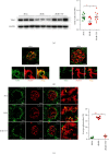
TSF protects podocyte-associated protein talin1 in db/db mice, which may involve increased expression of TRPC6 in podocytes. (a) Immunoblotting and quantitative analysis of talin1 (n = 3 experiments). (b) Immunofluorescence costaining (scale bars, 25 μm) of nephrin (red) and talin1 (green) showed that talin1 had a specific deletion in podocytes. (c) Immunofluorescence costaining (scale bars, 25 μm) of nephrin (red; red arrow) and TRPC6 (green) showed that increased expression of podocyte TRPC6 in db/db mice (yellow arrow) compared to db/m mice and db/db + TSF mice (n = 6). Fluorescent images were collected and assessed using a high-content screening system. The data were expressed as the mean ± SEM. #P < 0.05, ##P < 0.01 vs. db/m group; P < 0.05, ∗∗P < 0.01 vs. db/db group. Statistically analyzed via a one-way ANOVA with Dunnett's correction.
Figure 4
Figure 4
TSF attenuated the increased cell migration in AGEs-stimulated primary mice podocytes. (a, b) Wound healing assay and quantitative analysis using different doses of AGEs for 24 hours to observe the effect on primary mice podocytes (scale bars, 100 μm) (n = 3 experiments). (c, d) Viability of primary mice podocytes was determined with MTT assay (n = 3 experiments). (e, f) Wound healing assay and quantitative analysis using different doses of TSF for 24 hours (scale bars, 100 μm) (n = 3 experiments). The data were expressed as the mean ± SEM of three independent experiments performed in triplicate. #P < 0.05, ##P < 0.01 vs. BSA group; P < 0.05, ∗∗P < 0.01 vs. AGEs group. Statistically analyzed via a one-way ANOVA with Dunnett's correction.
Figure 5
Figure 5
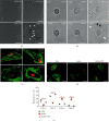
TSF inhibited actin-cytoskeletal disorganization in AGEs-stimulated primary mice podocytes. (a) Morphology of primary mice podocytes in wound healing assay (scale bars, 100 μm). (b) Time-lapse images of primary mice podocytes (scale bars, 50 μm). Lamellipodia are marked with white arrows and filopodia with black arrows. (c) Representative images of different types of phalloidin staining patterns observed in isolated control podocytes (scale bars, 25 μm). (d) Representative immunofluorescence stained with phalloidin and WT1 showed actin-cytoskeletal disorganization in AGEs-stimulated primary mice podocytes which could be relieved by TSF (scale bars, 50 μm). (e) Quantitative analysis of the percentage of four types of phalloidin staining patterns in (d) by a blinded observer (30 cells evaluated per experiment, n = 3 experiments). The data were expressed as the mean ± SEM of three independent experiments performed in triplicate. #P < 0.05, ##P < 0.01 vs. BSA group; P < 0.05, ∗∗P < 0.01 vs. AGEs group. Statistically analyzed via a one-way ANOVA with Dunnett's correction.
Figure 6
Figure 6
TSF improved TRPC6-dependent Ca2+ accumulation-mediated reduction of focal adhesions in AGEs-stimulated primary mice podocytes. (a) Effects of TSF or SAR7334 on intracellular Ca2+ level of AGEs-induced primary mice podocyte injury (n = 3 experiments). (b) Immunoblotting and quantitative analysis of talin1 (n = 3 experiments). (c) Immunoblotting and quantitative analysis of TRPC6 and integrin β1 (n = 3 experiments). (d) Representative immunofluorescence stained with talin1 (green) and paxillin (red) (scale bars, 50 μm). The data were expressed as the mean ± SEM of three independent experiments performed in triplicate. #P < 0.05, ##P < 0.01 vs. BSA group; P < 0.05, ∗∗P < 0.01 vs. AGEs group; ΔP < 0.05, ΔΔP < 0.01 vs. AGEs + TSF group. Statistically analyzed via a one-way ANOVA with Dunnett's correction.
Figure 7
Figure 7
TSF alleviated TRPC6-dependent Ca2+ accumulation-mediated actin-cytoskeletal disorganization in AGEs-stimulated primary mice podocytes. (a) Representative immunofluorescence stained with phalloidin and TRPC6 (scale bars, 25 μm). (b) Quantitative analysis of the percentage of four types of phalloidin staining patterns in (a) by an observer blinded to the group (30 cells evaluated per experiment, n = 3 experiments). The data were expressed as the mean ± SEM of three independent experiments performed in triplicate. #P < 0.05, ##P < 0.01 vs. BSA group; P < 0.05, ∗∗P < 0.01 vs. AGEs group; ΔP < 0.05, ΔΔP<0.01 vs. AGEs + TSF group. Statistically analyzed via a one-way ANOVA with Dunnett's correction.
Figure 8
Figure 8
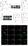
TSF alleviated NFATC2 nuclear translocation in AGEs-stimulated primary mice podocytes. (a) Representative immunofluorescence staining and quantitative analysis of NFATC2 (scale bars, 50 μm; 30 cells evaluated per experiment, n = 3 experiments). Nuclei were stained with DAPI. (b) Immunofluorescence staining for NFATC3 (scale bars, 50 μm). The data were expressed as the mean ± SEM. #P < 0.05, ##P < 0.01 vs. BSA group; P < 0.05, ∗∗P < 0.01 vs. AGEs group; ΔP < 0.05, ΔΔP < 0.01 vs. AGEs + TSF group. Statistically analyzed via a one-way ANOVA with Dunnett's correction.

Similar articles

Cited by

References

    1. Alicic R. Z., Rooney M. T., Tuttle K. R. Diabetic kidney disease: challenges, progress, and possibilities. Clinical Journal of the American Society of Nephrology: CJASN. 2017;12(12):2032–2045. doi: 10.2215/CJN.11491116. - DOI - PMC - PubMed
    1. Greka A., Mundel P. Cell biology and pathology of podocytes. Annual Review of Physiology. 2012;74(1):299–323. doi: 10.1146/annurev-physiol-020911-153238. - DOI - PMC - PubMed
    1. Fissell W. H., Miner J. H. What is the glomerular ultrafiltration barrier? Journal of Cell Biology. 2018;29(9):2262–2264. doi: 10.1681/asn.2018050490. - DOI - PMC - PubMed
    1. Faul C., Asanuma K., Yanagida-Asanuma E., Kim K., Mundel P. Actin up: regulation of podocyte structure and function by components of the actin cytoskeleton. Trends in Cell Biology. 2007;17(9):428–437. doi: 10.1016/j.tcb.2007.06.006. - DOI - PubMed
    1. Maezawa Y., Yokote K. Human glomerular transcriptome of diabetic kidneys: can the podocyte cytoskeleton be a therapeutic target? Journal of Diabetes Investigation. 2019;10(2):224–226. doi: 10.1111/jdi.12951. - DOI - PMC - PubMed

MeSH terms