

Immunohistochemical analysis of protein expression in formalin fixed paraffin embedded human intervertebral disc tissues
- PMID: 33015573
- PMCID: PMC7524243
- DOI: 10.1002/jsp2.1098
Immunohistochemical analysis of protein expression in formalin fixed paraffin embedded human intervertebral disc tissues
Abstract
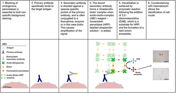
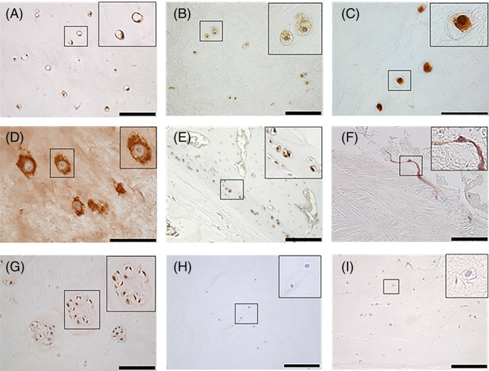
Immunohistochemistry (IHC) is a useful technique for the localization and semiquantification of protein expression within tissues. Adult human intervertebral disc (IVD) tissues contain a large amount of auto-fluorescence which often makes immunofluorescence techniques inappropriate on tissue samples but can be applied to isolated cell samples. Thus, IHC remains one of, if not the most common application for protein detection within IVD tissue. Immunostaining localizes antigen expression through specific epitope-antibody interactions. Within the field of IVD research, IHC is commonly used on fresh frozen and paraffin embedded tissues to elucidate the expression of antigens. Here, we discuss the principles of IHC applied to formalin fixed paraffin embedded IVD tissue and supply optimized protocols for antibodies used within our group to guide research within the IVD field.
Keywords: immunohistochemistry; intervertebral disc; protein localisation; protocol.
© 2020 The Authors. JOR Spine published by Wiley Periodicals LLC. on behalf of Orthopaedic Research Society.
Conflict of interest statement
There are no conflict of interest to declare.
Figures
References
-
- Fixation and fixatives (2)—Factors influencing chemical fixation, formaldehyde and glutaraldehyde: leica biosystems. https://www.leicabiosystems.com/knowledge-pathway/fixation-and-fixatives.... Accessed April 8, 2020.
-
- An introduction to decalcification: leica biosystems. https://www.leicabiosystems.com/knowledge-pathway/an-introduction-to-dec.... Accessed April 8, 2020.
-
- D'Amico F, Skarmoutsou E, Stivala F. State of the art in antigen retrieval for immunohistochemistry. J Immunol Methods. 2009;341:1‐18. - PubMed
-
- Shi SR, Key ME, Kalra KL. Antigen retrieval in formalin‐fixed, paraffin‐embedded tissues: an enhancement method for immunohistochemical staining based on microwave oven heating of tissue sections. J Histochem Cytochem. 1991;39:741‐748. - PubMed
Grants and funding
LinkOut - more resources
Full Text Sources
Other Literature Sources
