. 2020 Nov;1(7):e290-e299.
doi: 10.1016/S2666-5247(20)30144-0. Epub 2020 Sep 25.

Viral presence and immunopathology in patients with lethal COVID-19: a prospective autopsy cohort study

Affiliations

Viral presence and immunopathology in patients with lethal COVID-19: a prospective autopsy cohort study

Bernadette Schurink et al. Lancet Microbe. 2020 Nov.

Abstract

Background: Severe acute respiratory syndrome coronavirus 2 (SARS-CoV-2) targets multiple organs and causes severe coagulopathy. Histopathological organ changes might not only be attributable to a direct virus-induced effect, but also the immune response. The aims of this study were to assess the duration of viral presence, identify the extent of inflammatory response, and investigate the underlying cause of coagulopathy.

Methods: This prospective autopsy cohort study was done at Amsterdam University Medical Centers (UMC), the Netherlands. With informed consent from relatives, full body autopsy was done on 21 patients with COVID-19 for whom autopsy was requested between March 9 and May 18, 2020. In addition to histopathological evaluation of organ damage, the presence of SARS-CoV-2 nucleocapsid protein and the composition of the immune infiltrate and thrombi were assessed, and all were linked to disease course.

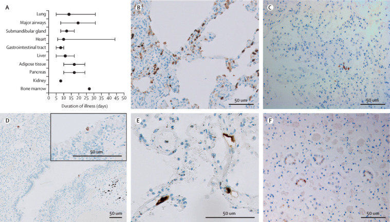
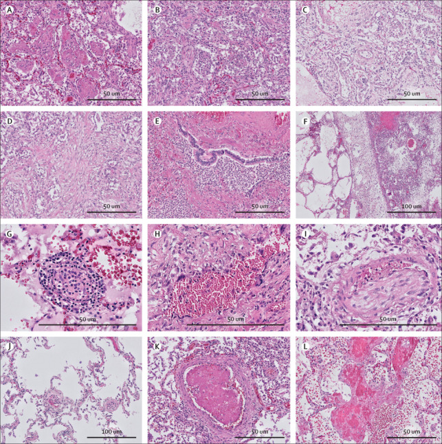
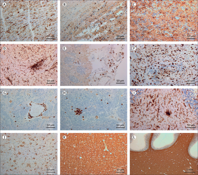
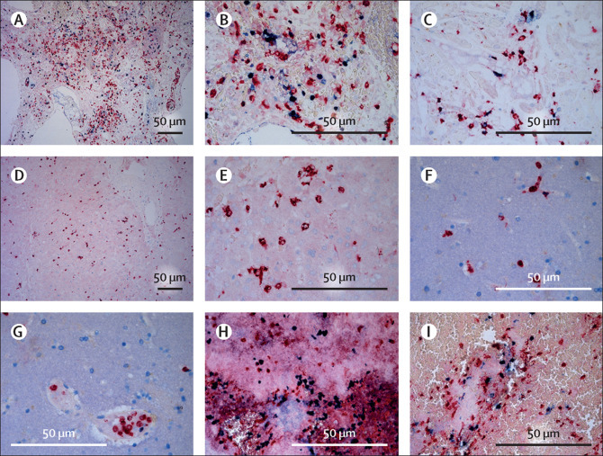
Findings: Our cohort (n=21) included 16 (76%) men, and median age was 68 years (range 41-78). Median disease course (time from onset of symptoms to death) was 22 days (range 5-44 days). In 11 patients tested for SARS-CoV-2 tropism, SARS-CoV-2 infected cells were present in multiple organs, most abundantly in the lungs, but presence in the lungs became sporadic with increased disease course. Other SARS-CoV-2-positive organs included the upper respiratory tract, heart, kidneys, and gastrointestinal tract. In histological analyses of organs (sampled from nine to 21 patients per organ), an extensive inflammatory response was present in the lungs, heart, liver, kidneys, and brain. In the brain, extensive inflammation was seen in the olfactory bulbs and medulla oblongata. Thrombi and neutrophilic plugs were present in the lungs, heart, kidneys, liver, spleen, and brain and were most frequently observed late in the disease course (15 patients with thrombi, median disease course 22 days [5-44]; ten patients with neutrophilic plugs, 21 days [5-44]). Neutrophilic plugs were observed in two forms: solely composed of neutrophils with neutrophil extracellular traps (NETs), or as aggregates of NETs and platelets..

Interpretation: In patients with lethal COVID-19, an extensive systemic inflammatory response was present, with a continued presence of neutrophils and NETs. However, SARS-CoV-2-infected cells were only sporadically present at late stages of COVID-19. This suggests a maladaptive immune response and substantiates the evidence for immunomodulation as a target in the treatment of severe COVID-19.

Funding: Amsterdam UMC Corona Research Fund.


Figures

Figure 1
Figure 1
SARS-CoV-2 tropism Two antibodies against SARS-CoV-2 nucleocapsid protein were used to detect infected cells. Staining of the same cell type by both antibodies was considered as positive immunoreactivity. (A) Median disease course per organ group with immunoreactivity for SARS-CoV-2. Error bars indicate the range. Adipose tissue consisted of mesocolic fat or omental fat (or both). The appendix (p 7)) shows SARS-CoV-2 positivity per organ per patient. (B) Stain against SARS-CoV-2 in the lung of a patient with mainly exudative diffuse alveolar damage and a disease course of 5 days. Immunoreactive cells were abundant (>10% infected cells per high-power field). Infected cells were pneumocytes along the alveolar walls, stromal cells in the septae, endothelial cells in the small blood vessels, and alveolar macrophages. (C) Stain against SARS-CoV-2 in the lung later in the disease course (patient with a disease course of 22 days) revealed only scattered immunoreactive cells, conceivably pneumocytes. (D) Stain against SARS-CoV-2 in the lung later in the disease course (patient with a disease course of 31 days) also showed immunopositivity in a respiratory cell lining a bronchiole. (E) Stain against SARS-CoV-2 in the lung early in the disease course (patient with a disease course of 5 days [also represented in part B]) showed immunopositive endothelial cells in septal capillaries. (F) Stain against SARS-CoV-2 in the kidney (patient with a disease course of 24 days) revealed immunoreactivity of the distal tubular epithelial cells. SARS-CoV-2=severe acute respiratory syndrome coronavirus 2.
Figure 2
Figure 2
Lung histopathology in COVID-19 Different phases of diffuse alveolar damage were identified in COVID-19 (haematoxylin and eosin stain). (A) Exudative pattern with intra-alveolar fibrin exudation. (B) Exudative pattern with desquamation with early fibroblastic proliferation. (C) Proliferative diffuse alveolar damage with fibroblastic proliferation in the alveoli, partially incorporated in the alveolar septa. (D) Fibrosing phase of alveolar damage with collagen deposition (pink) in the areas with fibroblastic proliferation. (E) Exudative bronchopneumonia with neutrophil granulocyte infiltration of bronchi and surrounding alveolar parenchyma. (F) Patchy distribution of the acute damage and prominent lymphatic stasis in the septa. (G) Endotheliitis of small vessels (<100 μm) with infiltration of the endothelium and vessel wall by lymphocytes and plasma cells. (H) Giant cell transformation of the endothelium in a patient with longstanding COVID-19 (disease course 30 days). (I) Chronic thromboembolic vasculopathy with an organised thrombus in an arteriole. (J) Patchy thrombi in microvessels (<70 μm) and segregation of thrombocytes and neutrophil granulocytes in the vessels in the spared lung parenchyma. (K) New-formed thrombus in an arteriole. (L) Focal necrosis of the alveolar septa with blood and fibrin exudation in the parenchyma.
Figure 3
Figure 3
Neuroinflammatory response to COVID-19 in the brain (A) Stain against HLA-DR in the bulbus olfactorius showed numerous activated microglia with enlarged cell bodies and thick cell processes longitudinally arranged. (B) Stain against CD3 in the bulbus olfactorius revealed T-cell extravasion into the parenchyma. (C) Stain against glial fibrillary acidic protein in the bulbus olfactorius showed reactive astrocytes in an anisomorphic arrangement. (D) Stain against HLA-DR in the medulla oblongata in the region of the nucleus of the tractus solitarius showed massive microglia activation with formation of a large cell aggregate (microglia nodule). (E) Stain against CD3 in the dorsal aspect of the medulla oblongata revealed T cells in the leptomeninges. (F) Stain against HLA-DR in the cervical spinal cord showed activated microglia with ameboid morphology also in this region. (G) Stain against CD3 in the medulla oblongata indicated a perivascular cuffing of T cells; some cells can also be seen in the surrounding parenchyma. (H) Stain against CD3 in the medulla oblongata confirmed presence of intraparenchymal T cells, aggregated in small nodules. (I) Stain against HLA-DR in the cerebellum showed numerous activated microglia with enlarged cell bodies and thick processes in the white matter of the basis of a folium; a small microglia nodule was also present. (J) Stain against HLA-DR of the nucleus dentatus revealed activated microglia also in the cerebellar structures of deep grey matter. (K) Stain against the major myelin protein, MBP in the frontal lobe revealed preservation of myelin in the cerebral white matter. (L) Stain against MBP in the cerebellum showed intact myelin in the deep hemispheric regions and the folia. MBP=myelin basic protein.
Figure 4
Figure 4
Presence of neutrophils and formation of neutrophil extracellular traps Tissue sections of patients who died of severe acute respiratory syndrome coronavirus 2 infection were stained for myeloperoxidase (red), as a marker of neutrophilic granulocytes, and citrullinated histone 3 (blue), as a marker of extracellular DNA traps and where extracellular co-localisation suggests trap formation by neutrophils. (A and B) Lung tissue showed abundant presence of neutrophils in both the lung vasculature and parenchyma with formation of neutrophil extracellular traps in a patient with a disease course of 8 days. (C) Heart tissue showed the presence of neutrophils in and surrounding cardiac vessels and in the cardiac parenchyma with formation of neutrophil extracellular traps in a patient with a disease course of 17 days. (D and E) Liver tissue showed the presence of neutrophils in the liver parenchyma but no extracellular traps in a patient with a disease course of 27 days. (F and G) Brain tissue showed the presence of neutrophils within the cerebral vasculature but no extracellular traps in a patient with a disease course of 8 days. (H and I) Thrombus in the main bronchus showed abundant presence of neutrophils with formation of neutrophil extracellular traps in a patient with a disease course of 8 days.

Comment in

References

    1. Menter T, Haslbauer JD, Nienhold R. Postmortem examination of COVID-19 patients reveals diffuse alveolar damage with severe capillary congestion and variegated findings in lungs and other organs suggesting vascular dysfunction. Histopathology. 2020;77:198–209. - PMC - PubMed
    1. Ackermann M, Verleden SE, Kuehnel M. Pulmonary vascular endothelialitis, thrombosis, and angiogenesis in COVID-19. N Engl J Med. 2020;383:120–128. - PMC - PubMed
    1. Zeng J-H, Liu Y-X, Yuan J. First case of COVID-19 complicated with fulminant myocarditis: a case report and insights. Infection. 2020 doi: 10.1007/s15010-020-01424-5. published online April 10. - DOI - PMC - PubMed
    1. Lax SF, Skok K, Zechner P. Pulmonary arterial thrombosis in COVID-19 with fatal outcome: results from a prospective, single-center, clinicopathologic case series. Ann Intern Med. 2020;173:350–361. - PMC - PubMed
    1. Wichmann D, Sperhake J-P, Lütgehetmann M. Autopsy findings and venous thromboembolism in patients with COVID-19: a prospective cohort study. Ann Intern Med. 2020;173:268–277. - PMC - PubMed

Publication types