Review
. 2021 Mar;17(1):41-47.
doi: 10.1007/s11302-020-09743-w. Epub 2020 Oct 5.

Nociceptive signaling of P2X receptors in chronic pain states

Affiliations
Review

Nociceptive signaling of P2X receptors in chronic pain states

Kazuhide Inoue. Purinergic Signal. 2021 Mar.

Abstract

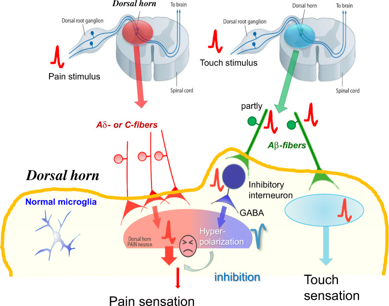
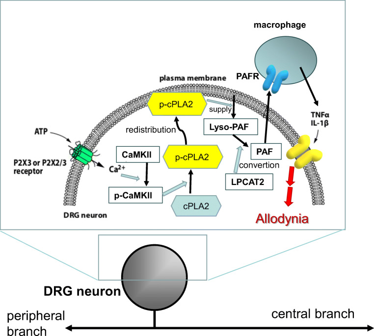
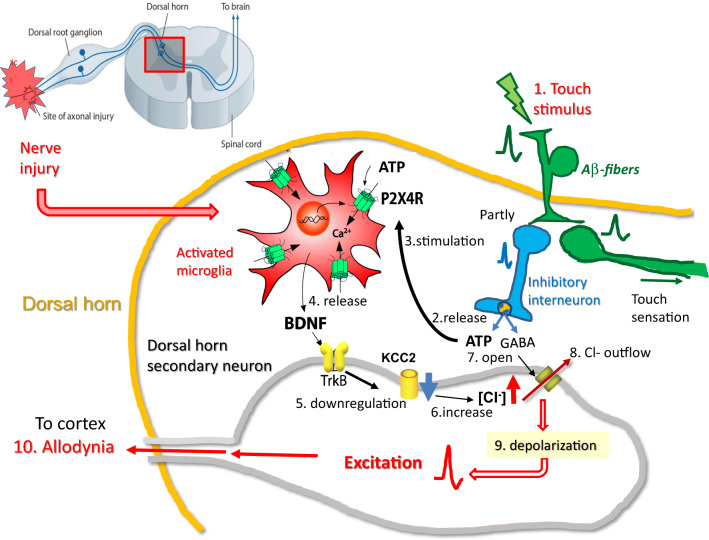
P2X3 monomeric receptors (P2X3Rs) and P2X2/3 heteromeric receptors (P2X2/3Rs) in primary sensory neurons and microglial P2X4 monomeric receptors (P2X4Rs) in the spinal dorsal horn (SDH) play important roles in neuropathic pain. In particular, P2X4R in the spinal microglia during peripheral nerve injury (PNI), experimental autoimmune neuritis, and herpes models are useful to explore the potential strategies for developing new drugs to treat neuropathic pain. Recently, novel P2X4 antagonists, NP-1815-PX and NC-2600, were developed, which demonstrated potent and specific inhibition against rodent and human P2X4Rs. The phase I study of NC-2600 has been completed, and no serious side effects were reported. The roles played by purinergic receptors in evoking neuropathic pain provide crucial insights into the pathogenesis of neuropathic pain.

Keywords: Microglia; Neuropathic pain; P2X2/3; P2X3; P2X4; Primary sensory neurons.


Conflict of interest statement

Kazuhide Inoue declares that he/she has no conflict of interest.

Figures

Fig. 1
Fig. 1
Pain signaling under normal conditions Painful stimuli evoke action potentials in the distal ends of C-fibers or Aδ-fibers of DRG neurons, and these signals are conducted to the central ends of these DRG neurons and transmitted to the secondary sensory neurons in the SDH. These signals finally reach to the sensory cortex resulting in pain sensation. Touch stimuli evoke action potentials in Aβ-fibers of DRG neurons, and these signals are transmitted to the sensory cortex, resulting in touch sensation. These action potentials can also be partially transmitted to the inhibitory interneurons in the SDH, resulting in the release of the inhibitory neurotransmitters, γ-aminobutyric acid (GABA), and glycine. GABA evokes the hyperpolarization of secondary neurons, which inhibits pain signaling.
Fig. 2
Fig. 2
Schema of the proposed mechanism for the P2X3R- and P2X2/3Rs-involved PAF/PAFR system-mediated tactile allodynia after PNI In DRG neurons, ATP stimulates P2X3Rs and P2X2/3Rs to increase internal concentration of Ca2+ after PNI, leading activation of Ca2+/calmodulin-dependent protein kinase II (CaMKII) which phosphorylates cPLA2. Phosphorylated cPLA2 (phospho-cPLA2) localizes in the vicinity of plasma membrane to cut out lyso-PAF from the cell membrane, which in turn converts into PAF by the lyso-PAF-acetyltransferase (LPCAT2). In macrophages, stimulation of PAFR by PAF may lead to produce and release of pro-inflammatory cytokines, TNFα and IL-1β. These cytokines may increase the excitability of DRG neurons that link to PNI-induced tactile allodynia
Fig.3
Fig.3
A hypothesis of the mechanism of allodynia involving microglial P2X4Rs PNI activates spinal microglia to overexpress P2X4Rs. In this pathological condition, touch stimuli (1) cause ATP and GABA release (2) from inhibitory interneurons of the DH. Released ATP stimulates (3) microglial P2X4Rs to secrete BDNF (4) which acts on secondary neurons to increase [Cl]i(6). Furthermore, released GABA affects the Cl channels of secondary neurons (7), leading to increase Cl outflow (8), resulting in depolarizing these neurons to evoke action potentials (9). These spikes reach the cortex and evoke pain (10). In this way, innocuous touch stimuli are mistakenly recognized as pain
Fig. 4
Fig. 4
Photos in memories with Geoffrey Burnstock

References

    1. Burnstock G. Purinergic P2 receptors as targets for novel analgesics. Pharmacol Ther. 2006;110:433–454. - PubMed
    1. Aldskogius H, Kozlova EN. Central neuron–glial and glial–glial interactions following axon injury. Prog Neurobiol. 1998;55:1–26. - PubMed
    1. Carson MJ. Microglia as liaisons between the immune and central nervous systems: functional implications for multiple sclerosis. Glia. 2002;40:218–231. - PMC - PubMed
    1. Eikelenboom P, Bate C, Van Gool WA, Hoozemans JJ, Rozemuller JM, Veerhuis R, Williams A. Neuroinflammation in Alzheimer’s disease and prion disease. Glia. 2002;40:232–239. - PubMed
    1. Colburn RW, Rickman AJ, Deleo JA. The effect of site and type of nerve injury on spinal glial activation and neuropathic pain behavior. Exp Neurol. 1999;157:289–304. - PubMed

Publication types

Substances

LinkOut - more resources